 Movie
Movie Controller

[English] 日本語
 Yorodumi
- PDB-2ef9: Structural and mechanistic changes along an engineered path from ... -
+ Open data

- Basic information
| Entry | Database: PDB / ID: 2ef9 | ||||||
|---|---|---|---|---|---|---|---|
| Title | Structural and mechanistic changes along an engineered path from metallo to non-metallo KDO8P synthase | ||||||
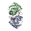
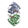
 Components Components | 2-dehydro-3-deoxyphosphooctonate aldolase | ||||||
 Keywords Keywords |  TRANSFERASE / KDO / KDO8P / KDO8PS / PEP / A5P TRANSFERASE / KDO / KDO8P / KDO8PS / PEP / A5P | ||||||
| Function / homology |  Function and homology information Function and homology informationmonosaccharide biosynthetic process / 3-deoxy-8-phosphooctulonate synthase / 3-deoxy-8-phosphooctulonate synthase activity / keto-3-deoxy-D-manno-octulosonic acid biosynthetic process / cytosolSimilarity search - Function | ||||||
| Biological species |   Aquifex aeolicus (bacteria) Aquifex aeolicus (bacteria) | ||||||
| Method | X-RAY DIFFRACTION / MOLECULAR REPLACEMENT / Resolution: 2 Å | ||||||
 Authors Authors | Kona, F. / Xu, X. / Martin, P. / Kuzmic, P. / Gatti, D.L. | ||||||
 Citation Citation | Journal: Biochemistry / Year: 2007 Title: Structural and Mechanistic Changes along an Engineered Path from Metallo to Nonmetallo 3-Deoxy-d-manno-octulosonate 8-Phosphate Synthases Authors: Kona, F. / Xu, X. / Martin, P. / Kuzmic, P. / Gatti, D.L. | ||||||
| History |
|
- Structure visualization
| Structure viewer | Molecule: MolmilJmol/JSmol |
|---|
- Downloads & links
-Download
| PDBx/mmCIF format | 2ef9.cif.gz | 123.2 KB | Display | PDBx/mmCIF format |
|---|---|---|---|---|
| PDB format | pdb2ef9.ent.gz | 95.5 KB | Display | PDB format |
| PDBx/mmJSON format | 2ef9.json.gz | Tree view | PDBx/mmJSON format | |
| Others |  Other downloads Other downloads |
-Validation report
| Arichive directory | https://data.pdbj.org/pub/pdb/validation_reports/ef/2ef9ftp://data.pdbj.org/pub/pdb/validation_reports/ef/2ef9 | HTTPS FTP |
|---|
-Related structure data
| Related structure data | 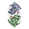 2nwrC  2nwsC 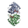 2nx1C  2nx3C  2nxgC 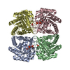 2nxhC 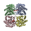 2nxiC 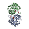 1fwnS C: citing same article ( S: Starting model for refinement |
|---|---|
| Similar structure data |


-Links
 PDBj
PDBj
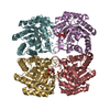
- Assembly
| Deposited unit | 
| ||||||||
|---|---|---|---|---|---|---|---|---|---|
| 1 | 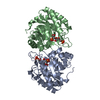
| ||||||||
| Unit cell |
|
-Components
| #1: Protein | Mass: 29772.434 Da / Num. of mol.: 2 / Mutation: P10M/C11N/S235P/Q237A Source method: isolated from a genetically manipulated source Source: (gene. exp.) Aquifex aeolicus (bacteria) / Production host: Escherichia coli (E. coli)References: UniProt: O66496, 3-deoxy-8-phosphooctulonate synthase#2: Chemical | Phosphoenolpyruvic acid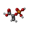  Mass: 168.042 Da / Num. of mol.: 2 / Source method: obtained synthetically / Formula: C3H5O6P Mass: 168.042 Da / Num. of mol.: 2 / Source method: obtained synthetically / Formula: C3H5O6P#3: Chemical |   Mass: 232.126 Da / Num. of mol.: 2 / Source method: obtained synthetically / Formula: C5H13O8P Mass: 232.126 Da / Num. of mol.: 2 / Source method: obtained synthetically / Formula: C5H13O8P#4: Water | ChemComp-HOH / | Water Mass: 18.015 Da / Num. of mol.: 402 / Source method: isolated from a natural source / Formula: H2O Mass: 18.015 Da / Num. of mol.: 402 / Source method: isolated from a natural source / Formula: H2O |
|---|
-Experimental details
-Experiment
| Experiment | Method: X-RAY DIFFRACTION / Number of used crystals: 1 |
|---|
- Sample preparation
| Crystal | Density Matthews: 2.77 Å3/Da / Density % sol: 55.57 % |
|---|
-Data collection
| Diffraction source | Source: ROTATING ANODE / Type: RIGAKU / Wavelength: 1.5418 Å |
|---|---|
| Radiation | Protocol: SINGLE WAVELENGTH / Monochromatic (M) / Laue (L): M / Scattering type: x-ray |
| Radiation wavelength | Wavelength: 1.5418 Å / Relative weight: 1 |
| Reflection | Resolution: 2→26.16 Å / Num. all: 45441 / Num. obs: 44624 / % possible obs: 0.982 % / Observed criterion σ(F): 0 / Observed criterion σ(I): 0 / Biso Wilson estimate: 13.2 Å2 |
- Processing
| Software | Name: CNS / Version: 1.1 / Classification: refinement | ||||||||||||||||||||||||||||||||||||||||||||||||||||||||||||||||||||||||||||||||
|---|---|---|---|---|---|---|---|---|---|---|---|---|---|---|---|---|---|---|---|---|---|---|---|---|---|---|---|---|---|---|---|---|---|---|---|---|---|---|---|---|---|---|---|---|---|---|---|---|---|---|---|---|---|---|---|---|---|---|---|---|---|---|---|---|---|---|---|---|---|---|---|---|---|---|---|---|---|---|---|---|---|
| Refinement | Method to determine structure: MOLECULAR REPLACEMENT Starting model: PDB Entry 1FWN Resolution: 2→26.16 Å / Rfactor Rfree error: 0.003 / Data cutoff high absF: 834328.8 / Data cutoff low absF: 0 / Isotropic thermal model: RESTRAINED / Cross valid method: THROUGHOUT / σ(F): 0
| ||||||||||||||||||||||||||||||||||||||||||||||||||||||||||||||||||||||||||||||||
| Solvent computation | Solvent model: FLAT MODEL / Bsol: 49.1537 Å2 / ksol: 0.338698 e/Å3 | ||||||||||||||||||||||||||||||||||||||||||||||||||||||||||||||||||||||||||||||||
| Displacement parameters | Biso mean: 26.3 Å2
| ||||||||||||||||||||||||||||||||||||||||||||||||||||||||||||||||||||||||||||||||
| Refine analyze |
| ||||||||||||||||||||||||||||||||||||||||||||||||||||||||||||||||||||||||||||||||
| Refinement step | Cycle: LAST / Resolution: 2→26.16 Å
| ||||||||||||||||||||||||||||||||||||||||||||||||||||||||||||||||||||||||||||||||
| Refine LS restraints |
| ||||||||||||||||||||||||||||||||||||||||||||||||||||||||||||||||||||||||||||||||
| LS refinement shell | Resolution: 2→2.13 Å / Rfactor Rfree error: 0.012 / Total num. of bins used: 6
| ||||||||||||||||||||||||||||||||||||||||||||||||||||||||||||||||||||||||||||||||
| Xplor file |
|
