 Movie
Movie Controller

[English] 日本語
 Yorodumi
- PDB-2ecq: Crystal Structure of T.th. HB8 O-acetylserine sulfhydrylase Compl... -
+ Open data

- Basic information
| Entry | Database: PDB / ID: 2ecq | ||||||
|---|---|---|---|---|---|---|---|
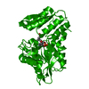
| Title | Crystal Structure of T.th. HB8 O-acetylserine sulfhydrylase Complexed with 3-Hydroxylactate | ||||||
 Components Components | O-acetylserine (Thiol)-lyase | ||||||
 Keywords Keywords |  TRANSFERASE / PLP-dependent TRANSFERASE / PLP-dependent | ||||||
| Function / homology |  Function and homology informationcysteine synthase / cysteine synthase activity / cysteine biosynthetic process from serine Function and homology informationcysteine synthase / cysteine synthase activity / cysteine biosynthetic process from serineSimilarity search - Function | ||||||
| Biological species |   Thermus thermophilus (bacteria) Thermus thermophilus (bacteria) | ||||||
| Method | X-RAY DIFFRACTION / MOLECULAR REPLACEMENT / Resolution: 1.9 Å | ||||||
 Authors Authors | Goto, M. | ||||||
 Citation Citation | Journal: to be published Title: Crystal Structure of T.th. HB8 O-acetylserine sulfhydrylase Complexed with 3-Hydroxylactate Authors: Goto, M. | ||||||
| History |
|
- Structure visualization
| Structure viewer | Molecule: MolmilJmol/JSmol |
|---|
- Downloads & links
-Download
| PDBx/mmCIF format | 2ecq.cif.gz | 79.3 KB | Display | PDBx/mmCIF format |
|---|---|---|---|---|
| PDB format | pdb2ecq.ent.gz | 57.6 KB | Display | PDB format |
| PDBx/mmJSON format | 2ecq.json.gz | Tree view | PDBx/mmJSON format | |
| Others |  Other downloads Other downloads |
-Validation report
| Arichive directory | https://data.pdbj.org/pub/pdb/validation_reports/ec/2ecqftp://data.pdbj.org/pub/pdb/validation_reports/ec/2ecq | HTTPS FTP |
|---|
-Related structure data
| Related structure data | 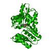 1ve1S S: Starting model for refinement |
|---|---|
| Similar structure data |


-Links
 PDBj
PDBj
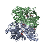
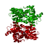
- Assembly
| Deposited unit | 
| ||||||||
|---|---|---|---|---|---|---|---|---|---|
| 1 | 
| ||||||||
| Unit cell |
|
-Components
| #1: Protein | Mass: 33065.094 Da / Num. of mol.: 1 Source method: isolated from a genetically manipulated source Source: (gene. exp.) Thermus thermophilus (bacteria) / Strain: HB8 / Plasmid: pET-20b / Production host: Escherichia coli (E. coli) / Strain (production host): BL21-CodonPlus(DE3)-RP / References: UniProt: Q5SLE6, cysteine synthase |
|---|---|
| #2: Chemical | ChemComp-PLP / Pyridoxal phosphate  Mass: 247.142 Da / Num. of mol.: 1 / Source method: obtained synthetically / Formula: C8H10NO6P Mass: 247.142 Da / Num. of mol.: 1 / Source method: obtained synthetically / Formula: C8H10NO6P |
| #3: Chemical | ChemComp-3HL / (  Mass: 104.105 Da / Num. of mol.: 1 / Source method: obtained synthetically / Formula: C4H8O3 Mass: 104.105 Da / Num. of mol.: 1 / Source method: obtained synthetically / Formula: C4H8O3 |
| #4: Water | ChemComp-HOH / Water Mass: 18.015 Da / Num. of mol.: 336 / Source method: isolated from a natural source / Formula: H2O Mass: 18.015 Da / Num. of mol.: 336 / Source method: isolated from a natural source / Formula: H2O |
-Experimental details
-Experiment
| Experiment | Method: X-RAY DIFFRACTION / Number of used crystals: 1 |
|---|
- Sample preparation
| Crystal | Density Matthews: 3.8 Å3/Da / Density % sol: 67.67 % |
|---|---|
| Crystal grow | Temperature: 293 K / Method: vapor diffusion, hanging drop / pH: 8.5 Details: Ammonium Sulfate, Tris-HCl, Glycerol, pH 8.5, VAPOR DIFFUSION, HANGING DROP, temperature 293K |
-Data collection
| Diffraction | Mean temperature: 100 K |
|---|---|
| Diffraction source | Source: ROTATING ANODE / Type: RIGAKU RU200 / Wavelength: 1.5418 Å |
| Detector | Type: RIGAKU RAXIS IV / Detector: IMAGE PLATE / Date: Mar 22, 2002 / Details: mirrors |
| Radiation | Protocol: SINGLE WAVELENGTH / Monochromatic (M) / Laue (L): M / Scattering type: x-ray |
| Radiation wavelength | Wavelength: 1.5418 Å / Relative weight: 1 |
| Reflection | Resolution: 1.9→45.26 Å / Num. all: 39981 / Num. obs: 37594 / % possible obs: 93.8 % / Biso Wilson estimate: 16.5 Å2 |
| Reflection shell | Resolution: 1.9→1.97 Å / % possible all: 79.3 |
- Processing
| Software |
| ||||||||||||||||||||||||||||||||||||
|---|---|---|---|---|---|---|---|---|---|---|---|---|---|---|---|---|---|---|---|---|---|---|---|---|---|---|---|---|---|---|---|---|---|---|---|---|---|
| Refinement | Method to determine structure: MOLECULAR REPLACEMENT Starting model: 1VE1 Resolution: 1.9→39.37 Å / Rfactor Rfree error: 0.003 / Data cutoff high absF: 1742599.05 / Data cutoff low absF: 0 / Isotropic thermal model: RESTRAINED / Cross valid method: THROUGHOUT / σ(F): 0
| ||||||||||||||||||||||||||||||||||||
| Solvent computation | Solvent model: FLAT MODEL / Bsol: 61.2874 Å2 / ksol: 0.385086 e/Å3 | ||||||||||||||||||||||||||||||||||||
| Displacement parameters | Biso mean: 20.4 Å2
| ||||||||||||||||||||||||||||||||||||
| Refine analyze |
| ||||||||||||||||||||||||||||||||||||
| Refinement step | Cycle: LAST / Resolution: 1.9→39.37 Å
| ||||||||||||||||||||||||||||||||||||
| Refine LS restraints |
| ||||||||||||||||||||||||||||||||||||
| LS refinement shell | Resolution: 1.9→2.02 Å / Rfactor Rfree error: 0.01 / Total num. of bins used: 6
| ||||||||||||||||||||||||||||||||||||
| Xplor file |
|
