 Movie
Movie Controller


+ Open data

- Basic information
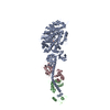
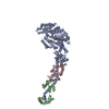
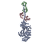
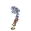
| Entry | Database: PDB / ID: 2ec6 | ||||||
|---|---|---|---|---|---|---|---|
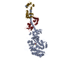
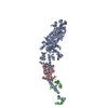
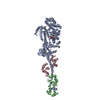
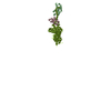
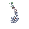
| Title | Placopecten Striated Muscle Myosin II | ||||||
 Components Components |
| ||||||
 Keywords Keywords |  CONTRACTILE PROTEIN / muscle / rigor-like / actin binding CONTRACTILE PROTEIN / muscle / rigor-like / actin binding | ||||||
| Function / homology |  Function and homology informationmyosin complex / myofibril / cytoskeletal motor activity / actin filament binding / calcium ion binding / ATP binding Function and homology informationmyosin complex / myofibril / cytoskeletal motor activity / actin filament binding / calcium ion binding / ATP bindingSimilarity search - Function | ||||||
| Biological species |  Placopecten magellanicus (sea scallop) Placopecten magellanicus (sea scallop) | ||||||
| Method | X-RAY DIFFRACTION / SYNCHROTRON / MOLECULAR REPLACEMENT / Resolution: 3.25 Å | ||||||
 Authors Authors | Yang, Y. / Brown, J. / Samudrala, G. / Reutzel, R. / Szent-Gyorgyi, A. | ||||||
 Citation Citation | Journal: Structure / Year: 2007 Title: Rigor-like structures from muscle myosins reveal key mechanical elements in the transduction pathways of this allosteric motor. Authors: Yang, Y. / Gourinath, S. / Kovacs, M. / Nyitray, L. / Reutzel, R. / Himmel, D.M. / O'Neall-Hennessey, E. / Reshetnikova, L. / Szent-Gyorgyi, A.G. / Brown, J.H. / Cohen, C. | ||||||
| History |
|
- Structure visualization
| Structure viewer | Molecule: MolmilJmol/JSmol |
|---|
- Downloads & links
-Download
| PDBx/mmCIF format | 2ec6.cif.gz | 225.7 KB | Display | PDBx/mmCIF format |
|---|---|---|---|---|
| PDB format | pdb2ec6.ent.gz | 180.2 KB | Display | PDB format |
| PDBx/mmJSON format | 2ec6.json.gz | Tree view | PDBx/mmJSON format | |
| Others |  Other downloads Other downloads |
-Validation report
| Arichive directory | https://data.pdbj.org/pub/pdb/validation_reports/ec/2ec6ftp://data.pdbj.org/pub/pdb/validation_reports/ec/2ec6 | HTTPS FTP |
|---|
-Related structure data
| Related structure data |  2os8C  2otgC  3i5fC  3i5gC  3i5hC  3i5iC C: citing same article ( |
|---|---|
| Similar structure data |

-Links
 PDBj
PDBj


- Assembly
| Deposited unit | 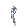
| ||||||||
|---|---|---|---|---|---|---|---|---|---|
| 1 |
| ||||||||
| Unit cell |
|
-Components
| #1: Protein | Myosin Mass: 95832.875 Da / Num. of mol.: 1 / Fragment: residues 1-838 Source method: isolated from a genetically manipulated source Details: The source was obtained from the Marine Biological Laboratory (MBL) Source: (gene. exp.) Placopecten magellanicus (sea scallop) / References: UniProt: Q26079 |
|---|---|
| #2: Protein | Mass: 15354.388 Da / Num. of mol.: 1 / Fragment: residues 23-156 / Mutation: Y84F, T151A Source method: isolated from a genetically manipulated source Details: The source was obtained from the Marine Biological Laboratory (MBL) Source: (gene. exp.) Placopecten magellanicus (sea scallop) / References: UniProt: Q26069 |
| #3: Protein | Mass: 17620.557 Da / Num. of mol.: 1 Source method: isolated from a genetically manipulated source Details: The source was obtained from the Marine Biological Laboratory (MBL) Source: (gene. exp.) Placopecten magellanicus (sea scallop) / References: UniProt: Q26066 |
| #4: Chemical | ChemComp-CA /   Mass: 40.078 Da / Num. of mol.: 1 / Source method: obtained synthetically / Formula: Ca Mass: 40.078 Da / Num. of mol.: 1 / Source method: obtained synthetically / Formula: Ca |
| #5: Water | ChemComp-HOH / Water Mass: 18.015 Da / Num. of mol.: 31 / Source method: isolated from a natural source / Formula: H2O Mass: 18.015 Da / Num. of mol.: 31 / Source method: isolated from a natural source / Formula: H2O |
-Experimental details
-Experiment
| Experiment | Method: X-RAY DIFFRACTION / Number of used crystals: 1 |
|---|
- Sample preparation
| Crystal | Density Matthews: 2.56 Å3/Da / Density % sol: 52.04 % |
|---|---|
| Crystal grow | Temperature: 277 K / Method: vapor diffusion, hanging drop / pH: 7.5 Details: 90mM CaCl2, 8.5% PEG 6K, 100mM HEPES, pH 7.5, VAPOR DIFFUSION, HANGING DROP, temperature 277K |
-Data collection
| Diffraction source | Source: SYNCHROTRON / Site: CHESS  / Beamline: A1 / Beamline: A1 |
|---|---|
| Detector | Type: ADSC QUANTUM 315 / Detector: CCD |
| Radiation | Protocol: SINGLE WAVELENGTH / Monochromatic (M) / Laue (L): M / Scattering type: x-ray |
| Radiation wavelength | Relative weight: 1 |
| Reflection | Resolution: 3.25→20 Å / Num. all: 21051 / Num. obs: 18825 / % possible obs: 94.7 % / Observed criterion σ(F): 1.5 / Observed criterion σ(I): 2 / Redundancy: 2.8 % / Rmerge(I) obs: 0.073 / Net I/σ(I): 12.4 |
| Reflection shell | Resolution: 3.25→20 Å / Redundancy: 2.6 % / Rmerge(I) obs: 0.458 / Mean I/σ(I) obs: 2 / Num. unique all: 1852 / Rsym value: 0.458 / % possible all: 87.8 |
- Processing
| Software |
| |||||||||||||||||||||||||
|---|---|---|---|---|---|---|---|---|---|---|---|---|---|---|---|---|---|---|---|---|---|---|---|---|---|---|
| Refinement | Method to determine structure: MOLECULAR REPLACEMENT / Resolution: 3.25→20 Å / σ(F): 1.5 / σ(I): 2 / Stereochemistry target values: Engh & Huber
| |||||||||||||||||||||||||
| Displacement parameters | Biso mean: 39.7 Å2 | |||||||||||||||||||||||||
| Refinement step | Cycle: LAST / Resolution: 3.25→20 Å
|
