 Movie
Movie Controller


+ Open data

- Basic information
| Entry | Database: PDB / ID: 2eb4 | ||||||
|---|---|---|---|---|---|---|---|
| Title | Crystal structure of apo-HpcG | ||||||
 Components Components | 2-oxo-hept-3-ene-1,7-dioate hydratase | ||||||
 Keywords Keywords |  LYASE / hydratase LYASE / hydratase | ||||||
| Function / homology |  Function and homology information2-hydroxyhexa-2,4-dienoate hydratase / 2-oxo-hept-3-ene-1,7-dioate hydratase activity / 2-hydroxyhexa-2,4-dienoate hydratase activity / 2-oxo-3-hexenedioate decarboxylase / 4-oxalocrotonate decarboxylase activity / 2-oxopent-4-enoate hydratase / 2-oxopent-4-enoate hydratase activity / : / metal ion binding Function and homology information2-hydroxyhexa-2,4-dienoate hydratase / 2-oxo-hept-3-ene-1,7-dioate hydratase activity / 2-hydroxyhexa-2,4-dienoate hydratase activity / 2-oxo-3-hexenedioate decarboxylase / 4-oxalocrotonate decarboxylase activity / 2-oxopent-4-enoate hydratase / 2-oxopent-4-enoate hydratase activity / : / metal ion bindingSimilarity search - Function | ||||||
| Biological species |  Escherichia coli (E. coli) Escherichia coli (E. coli) | ||||||
| Method | X-RAY DIFFRACTION / SYNCHROTRON / SAD / Resolution: 1.6 Å | ||||||
 Authors Authors | Izumi, A. / Rea, D. / Adachi, T. / Unzai, S. / Park, S.Y. / Roper, D.I. / Tame, J.R.H. | ||||||
 Citation Citation | Journal: J.Mol.Biol. / Year: 2007 Title: Structure and Mechanism of HpcG, a Hydratase in the Homoprotocatechuate Degradation Pathway of Escherichia coli Authors: Izumi, A. / Rea, D. / Adachi, T. / Unzai, S. / Park, S.Y. / Roper, D.I. / Tame, J.R.H. | ||||||
| History |
|
- Structure visualization
| Structure viewer | Molecule: MolmilJmol/JSmol |
|---|
- Downloads & links
-Download
| PDBx/mmCIF format | 2eb4.cif.gz | 279 KB | Display | PDBx/mmCIF format |
|---|---|---|---|---|
| PDB format | pdb2eb4.ent.gz | 225.4 KB | Display | PDB format |
| PDBx/mmJSON format | 2eb4.json.gz | Tree view | PDBx/mmJSON format | |
| Others |  Other downloads Other downloads |
-Validation report
| Arichive directory | https://data.pdbj.org/pub/pdb/validation_reports/eb/2eb4ftp://data.pdbj.org/pub/pdb/validation_reports/eb/2eb4 | HTTPS FTP |
|---|
-Related structure data


-Links
 PDBj
PDBj
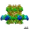
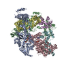
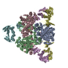
- Assembly
| Deposited unit | 
| |||||||||||||||||||||||||||||||||||||||||||||||||||||||||||||||||||||||||||||
|---|---|---|---|---|---|---|---|---|---|---|---|---|---|---|---|---|---|---|---|---|---|---|---|---|---|---|---|---|---|---|---|---|---|---|---|---|---|---|---|---|---|---|---|---|---|---|---|---|---|---|---|---|---|---|---|---|---|---|---|---|---|---|---|---|---|---|---|---|---|---|---|---|---|---|---|---|---|---|
| 1 | 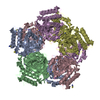
| |||||||||||||||||||||||||||||||||||||||||||||||||||||||||||||||||||||||||||||
| Unit cell |
| |||||||||||||||||||||||||||||||||||||||||||||||||||||||||||||||||||||||||||||
| Noncrystallographic symmetry (NCS) | NCS domain:
NCS domain segments: Component-ID: 1 / Beg auth comp-ID: MET / Beg label comp-ID: MET / End auth comp-ID: HIS / End label comp-ID: HIS / Auth seq-ID: 1 - 60 / Label seq-ID: 1 - 60
NCS ensembles :
|
-Components
| #1: Protein | Mass: 29750.828 Da / Num. of mol.: 5 Source method: isolated from a genetically manipulated source Source: (gene. exp.) Escherichia coli (E. coli) / Strain: strain C / Gene: hpcG / Plasmid: pET21 / Species (production host): Escherichia coli / Production host: Escherichia coli BL21(DE3) (bacteria) / Strain (production host): BL21(DE3)References: UniProt: Q46982, Lyases; Carbon-oxygen lyases; Hydro-lyases#2: Chemical | ChemComp-SCN / Thiocyanate  Mass: 58.082 Da / Num. of mol.: 9 / Source method: obtained synthetically / Formula: CNS Mass: 58.082 Da / Num. of mol.: 9 / Source method: obtained synthetically / Formula: CNS#3: Chemical | ChemComp-NA /   Mass: 22.990 Da / Num. of mol.: 5 / Source method: obtained synthetically / Formula: Na Mass: 22.990 Da / Num. of mol.: 5 / Source method: obtained synthetically / Formula: Na#4: Water | ChemComp-HOH / | Water Mass: 18.015 Da / Num. of mol.: 1008 / Source method: isolated from a natural source / Formula: H2O Mass: 18.015 Da / Num. of mol.: 1008 / Source method: isolated from a natural source / Formula: H2O |
|---|
-Experimental details
-Experiment
| Experiment | Method: X-RAY DIFFRACTION / Number of used crystals: 1 |
|---|
- Sample preparation
| Crystal | Density Matthews: 3.03 Å3/Da / Density % sol: 59.39 % |
|---|---|
| Crystal grow | Temperature: 293 K / Method: vapor diffusion, sitting drop / pH: 6 Details: 7% PEG 12000, 0.1M MES, 0.7M potassium thiocyanate, 7% PEG 550 MME, 10mM DTT, pH 6.0, VAPOR DIFFUSION, SITTING DROP, temperature 293K |
-Data collection
| Diffraction | Mean temperature: 100 K |
|---|---|
| Diffraction source | Source: SYNCHROTRON / Site: SPring-8  / Beamline: BL38B1 / Wavelength: 1 Å / Beamline: BL38B1 / Wavelength: 1 Å |
| Detector | Type: RIGAKU JUPITER 210 / Detector: CCD / Date: Oct 14, 2006 |
| Radiation | Monochromator: Si / Protocol: SINGLE WAVELENGTH / Monochromatic (M) / Laue (L): M / Scattering type: x-ray |
| Radiation wavelength | Wavelength: 1 Å / Relative weight: 1 |
| Reflection | Resolution: 1.6→50 Å / Num. obs: 223282 / % possible obs: 93.3 % / Observed criterion σ(F): 0 / Observed criterion σ(I): 0 / Redundancy: 6.2 % / Rmerge(I) obs: 0.069 / Net I/σ(I): 17.9 |
| Reflection shell | Resolution: 1.6→1.66 Å / Redundancy: 2.1 % / Rmerge(I) obs: 0.406 / % possible all: 68.4 |
- Processing
| Software |
| ||||||||||||||||||||||||||||||||||||||||||||||||||||||||||||||||||||||||||||||||||||||||||||||||||||||
|---|---|---|---|---|---|---|---|---|---|---|---|---|---|---|---|---|---|---|---|---|---|---|---|---|---|---|---|---|---|---|---|---|---|---|---|---|---|---|---|---|---|---|---|---|---|---|---|---|---|---|---|---|---|---|---|---|---|---|---|---|---|---|---|---|---|---|---|---|---|---|---|---|---|---|---|---|---|---|---|---|---|---|---|---|---|---|---|---|---|---|---|---|---|---|---|---|---|---|---|---|---|---|---|
| Refinement | Method to determine structure: SAD / Resolution: 1.6→20 Å / Cor.coef. Fo:Fc: 0.959 / Cor.coef. Fo:Fc free: 0.948 / SU B: 1.827 / SU ML: 0.064 / Cross valid method: THROUGHOUT / ESU R: 0.089 / ESU R Free: 0.088 / Stereochemistry target values: MAXIMUM LIKELIHOOD
| ||||||||||||||||||||||||||||||||||||||||||||||||||||||||||||||||||||||||||||||||||||||||||||||||||||||
| Solvent computation | Ion probe radii: 0.8 Å / Shrinkage radii: 0.8 Å / VDW probe radii: 1.4 Å / Solvent model: MASK | ||||||||||||||||||||||||||||||||||||||||||||||||||||||||||||||||||||||||||||||||||||||||||||||||||||||
| Displacement parameters | Biso mean: 26.347 Å2
| ||||||||||||||||||||||||||||||||||||||||||||||||||||||||||||||||||||||||||||||||||||||||||||||||||||||
| Refinement step | Cycle: LAST / Resolution: 1.6→20 Å
| ||||||||||||||||||||||||||||||||||||||||||||||||||||||||||||||||||||||||||||||||||||||||||||||||||||||
| Refine LS restraints |
| ||||||||||||||||||||||||||||||||||||||||||||||||||||||||||||||||||||||||||||||||||||||||||||||||||||||
| Refine LS restraints NCS | Dom-ID: 1 / Refine-ID: X-RAY DIFFRACTION
| ||||||||||||||||||||||||||||||||||||||||||||||||||||||||||||||||||||||||||||||||||||||||||||||||||||||
| LS refinement shell | Resolution: 1.599→1.64 Å / Total num. of bins used: 20
|
