 Movie
Movie Controller

[English] 日本語
 Yorodumi
- PDB-2e54: Crystal structure of acetylornithine aminotransferase from Thermo... -
+ Open data

- Basic information
| Entry | Database: PDB / ID: 2.0E+54 | ||||||
|---|---|---|---|---|---|---|---|
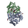
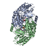
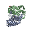
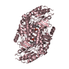
| Title | Crystal structure of acetylornithine aminotransferase from Thermotoga maritima | ||||||
 Components Components | Acetylornithine aminotransferase | ||||||
 Keywords Keywords |  TRANSFERASE / argD / Structural Genomics / NPPSFA / National Project on Protein Structural and Functional Analyses / RIKEN Structural Genomics/Proteomics Initiative / RSGI TRANSFERASE / argD / Structural Genomics / NPPSFA / National Project on Protein Structural and Functional Analyses / RIKEN Structural Genomics/Proteomics Initiative / RSGI | ||||||
| Function / homology |  Function and homology informationacetylornithine transaminase / N2-acetyl-L-ornithine:2-oxoglutarate 5-aminotransferase activity / arginine biosynthetic process / pyridoxal phosphate binding / identical protein binding / cytoplasm Function and homology informationacetylornithine transaminase / N2-acetyl-L-ornithine:2-oxoglutarate 5-aminotransferase activity / arginine biosynthetic process / pyridoxal phosphate binding / identical protein binding / cytoplasmSimilarity search - Function | ||||||
| Biological species |   Thermotoga maritima (bacteria) Thermotoga maritima (bacteria) | ||||||
| Method | X-RAY DIFFRACTION / SYNCHROTRON / MOLECULAR REPLACEMENT / Resolution: 1.5 Å | ||||||
 Authors Authors | Mizutani, H. / Kunishima, N. / RIKEN Structural Genomics/Proteomics Initiative (RSGI) | ||||||
 Citation Citation | Journal: To be Published Title: Crystal structure of acetylornithine aminotransferase from Thermotoga maritima Authors: Mizutani, H. / Kunishima, N. | ||||||
| History |
|
- Structure visualization
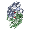
| Structure viewer | Molecule: MolmilJmol/JSmol |
|---|
- Downloads & links
-Download
| PDBx/mmCIF format | 2e54.cif.gz | 102.7 KB | Display | PDBx/mmCIF format |
|---|---|---|---|---|
| PDB format | pdb2e54.ent.gz | 76.2 KB | Display | PDB format |
| PDBx/mmJSON format | 2e54.json.gz | Tree view | PDBx/mmJSON format | |
| Others |  Other downloads Other downloads |
-Validation report
| Arichive directory | https://data.pdbj.org/pub/pdb/validation_reports/e5/2e54ftp://data.pdbj.org/pub/pdb/validation_reports/e5/2e54 | HTTPS FTP |
|---|
-Related structure data
| Related structure data | 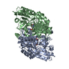 1vefS S: Starting model for refinement |
|---|---|
| Similar structure data | |
| Other databases |


-Links
 PDBj
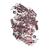
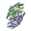
PDBj- Assembly
| Deposited unit | 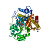
| |||||||||
|---|---|---|---|---|---|---|---|---|---|---|
| 1 | 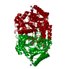
| |||||||||
| Unit cell |
| |||||||||
| Components on special symmetry positions |
| |||||||||
| Details | The biological assembly is a dimer generated by the operations: -x+1, -y, z+1. |
-Components
| #1: Protein | Mass: 42937.129 Da / Num. of mol.: 1 Source method: isolated from a genetically manipulated source Source: (gene. exp.) Thermotoga maritima (bacteria) / Strain: MSB8 / Plasmid: pET21a / Production host: Escherichia coli (E. coli) / Strain (production host): BL21-CodonPlus(DE3)-RIL / References: UniProt: Q9X2A5, acetylornithine transaminase | ||||||
|---|---|---|---|---|---|---|---|
| #2: Chemical |   Mass: 22.990 Da / Num. of mol.: 2 / Source method: obtained synthetically / Formula: Na Mass: 22.990 Da / Num. of mol.: 2 / Source method: obtained synthetically / Formula: Na#3: Chemical | ChemComp-PLP / | Pyridoxal phosphate  Mass: 247.142 Da / Num. of mol.: 1 / Source method: obtained synthetically / Formula: C8H10NO6P Mass: 247.142 Da / Num. of mol.: 1 / Source method: obtained synthetically / Formula: C8H10NO6P#4: Chemical | Ethylene glycol  Mass: 62.068 Da / Num. of mol.: 2 / Source method: obtained synthetically / Formula: C2H6O2 Mass: 62.068 Da / Num. of mol.: 2 / Source method: obtained synthetically / Formula: C2H6O2#5: Water | ChemComp-HOH / | Water Mass: 18.015 Da / Num. of mol.: 516 / Source method: isolated from a natural source / Formula: H2O Mass: 18.015 Da / Num. of mol.: 516 / Source method: isolated from a natural source / Formula: H2O |
-Experimental details
-Experiment
| Experiment | Method: X-RAY DIFFRACTION / Number of used crystals: 1 |
|---|
- Sample preparation
| Crystal | Density Matthews: 2.37 Å3/Da / Density % sol: 48.19 % |
|---|---|
| Crystal grow | Temperature: 295 K / Method: microbatch / pH: 8.5 Details: 0.2M NaCl, 0.1M Tris-HCl, 25%(w/v) PEG 3350, pH 8.5, microbatch, temperature 295K |
-Data collection
| Diffraction | Mean temperature: 100 K |
|---|---|
| Diffraction source | Source: SYNCHROTRON / Site: SPring-8  / Beamline: BL26B1 / Wavelength: 1 Å / Beamline: BL26B1 / Wavelength: 1 Å |
| Detector | Type: RIGAKU RAXIS V / Detector: IMAGE PLATE / Date: Oct 16, 2006 |
| Radiation | Monochromator: bending magnet / Protocol: SINGLE WAVELENGTH / Monochromatic (M) / Laue (L): M / Scattering type: x-ray |
| Radiation wavelength | Wavelength: 1 Å / Relative weight: 1 |
| Reflection | Resolution: 1.5→30 Å / Num. all: 66199 / Num. obs: 66199 / % possible obs: 99.9 % / Observed criterion σ(F): 0 / Observed criterion σ(I): 0 / Redundancy: 7 % / Biso Wilson estimate: 21.1 Å2 / Rmerge(I) obs: 0.066 / Rsym value: 0.063 / Net I/σ(I): 14.8 |
| Reflection shell | Resolution: 1.5→1.55 Å / Redundancy: 6.9 % / Rmerge(I) obs: 0.364 / Mean I/σ(I) obs: 5.35 / Num. unique all: 6498 / Rsym value: 0.34 / % possible all: 100 |
- Processing
| Software |
| ||||||||||||||||||||||||||||||||||||||||||||||||||||||||||||||||||||||||||||||||
|---|---|---|---|---|---|---|---|---|---|---|---|---|---|---|---|---|---|---|---|---|---|---|---|---|---|---|---|---|---|---|---|---|---|---|---|---|---|---|---|---|---|---|---|---|---|---|---|---|---|---|---|---|---|---|---|---|---|---|---|---|---|---|---|---|---|---|---|---|---|---|---|---|---|---|---|---|---|---|---|---|---|
| Refinement | Method to determine structure: MOLECULAR REPLACEMENT Starting model: PDB ENTRY 1VEF Resolution: 1.5→30 Å / Rfactor Rfree error: 0.004 / Data cutoff high absF: 1368180.57 / Data cutoff low absF: 0 / Isotropic thermal model: RESTRAINED / Cross valid method: THROUGHOUT / σ(F): 0 / Stereochemistry target values: Engh & Huber
| ||||||||||||||||||||||||||||||||||||||||||||||||||||||||||||||||||||||||||||||||
| Solvent computation | Solvent model: FLAT MODEL / Bsol: 65.4862 Å2 / ksol: 0.387397 e/Å3 | ||||||||||||||||||||||||||||||||||||||||||||||||||||||||||||||||||||||||||||||||
| Displacement parameters | Biso mean: 20.8 Å2
| ||||||||||||||||||||||||||||||||||||||||||||||||||||||||||||||||||||||||||||||||
| Refine analyze |
| ||||||||||||||||||||||||||||||||||||||||||||||||||||||||||||||||||||||||||||||||
| Refinement step | Cycle: LAST / Resolution: 1.5→30 Å
| ||||||||||||||||||||||||||||||||||||||||||||||||||||||||||||||||||||||||||||||||
| Refine LS restraints |
| ||||||||||||||||||||||||||||||||||||||||||||||||||||||||||||||||||||||||||||||||
| LS refinement shell | Resolution: 1.5→1.59 Å / Rfactor Rfree error: 0.011 / Total num. of bins used: 6
| ||||||||||||||||||||||||||||||||||||||||||||||||||||||||||||||||||||||||||||||||
| Xplor file |
|
