 Movie
Movie Controller

[English] 日本語
 Yorodumi
- PDB-2e32: Structural basis for selection of glycosylated substrate by SCFFb... -
+ Open data

- Basic information
| Entry | Database: PDB / ID: 2.0E+32 | ||||||
|---|---|---|---|---|---|---|---|
| Title | Structural basis for selection of glycosylated substrate by SCFFbs1 ubiquitin ligase | ||||||
 Components Components |
| ||||||
 Keywords Keywords |  LIGASE / ubiquitin / SCF / ubiquitin ligase / fbs1 LIGASE / ubiquitin / SCF / ubiquitin ligase / fbs1 | ||||||
| Function / homology |  Function and homology informationextrinsic component of postsynaptic membrane / denatured protein binding / regulation of protein catabolic process at postsynapse, modulating synaptic transmission / glycoprotein catabolic process / F-box domain binding / PcG protein complex / Neddylation / Antigen processing: Ubiquitination & Proteasome degradation / Cul7-RING ubiquitin ligase complex / positive regulation of ubiquitin protein ligase activity ...extrinsic component of postsynaptic membrane / denatured protein binding / regulation of protein catabolic process at postsynapse, modulating synaptic transmission / glycoprotein catabolic process / F-box domain binding / PcG protein complex / Neddylation / Antigen processing: Ubiquitination & Proteasome degradation / Cul7-RING ubiquitin ligase complex / positive regulation of ubiquitin protein ligase activity / maintenance of protein location in nucleus / Loss of Function of FBXW7 in Cancer and NOTCH1 Signaling / SCF-dependent proteasomal ubiquitin-dependent protein catabolic process / SCF ubiquitin ligase complex / ubiquitin ligase complex scaffold activity / Prolactin receptor signaling / protein monoubiquitination / cullin family protein binding / ubiquitin-like ligase-substrate adaptor activity / regulation of protein ubiquitination / protein K48-linked ubiquitination / Nuclear events stimulated by ALK signaling in cancer / : / Regulation of BACH1 activity / MAP3K8 (TPL2)-dependent MAPK1/3 activation / SCF-beta-TrCP mediated degradation of Emi1 / NIK-->noncanonical NF-kB signaling / molecular function activator activity / Vpu mediated degradation of CD4 / Dectin-1 mediated noncanonical NF-kB signaling / Degradation of GLI1 by the proteasome / Activation of NF-kappaB in B cells / Negative regulation of NOTCH4 signaling / Iron uptake and transport / GSK3B and BTRC:CUL1-mediated-degradation of NFE2L2 / Degradation of GLI2 by the proteasome / GLI3 is processed to GLI3R by the proteasome / FBXL7 down-regulates AURKA during mitotic entry and in early mitosis / Degradation of beta-catenin by the destruction complex / NOTCH1 Intracellular Domain Regulates Transcription / CLEC7A (Dectin-1) signaling / SCF(Skp2)-mediated degradation of p27/p21 / beta-catenin binding / Constitutive Signaling by NOTCH1 PEST Domain Mutants / Constitutive Signaling by NOTCH1 HD+PEST Domain Mutants / FCERI mediated NF-kB activation / Interleukin-1 signaling / protein polyubiquitination / Orc1 removal from chromatin / Regulation of RUNX2 expression and activity / Cyclin D associated events in G1 / Regulation of PLK1 Activity at G2/M Transition / Antigen processing: Ubiquitination & Proteasome degradation / Circadian Clock / Downstream TCR signaling / Neddylation / amyloid-beta binding / ubiquitin-dependent protein catabolic process / carbohydrate binding / proteasome-mediated ubiquitin-dependent protein catabolic process / dendritic spine / protein ubiquitination / chromatin remodeling / protein domain specific binding / negative regulation of cell population proliferation / centrosome / glutamatergic synapse / endoplasmic reticulum / nucleoplasm / nucleus / cytosol / cytoplasm Function and homology informationextrinsic component of postsynaptic membrane / denatured protein binding / regulation of protein catabolic process at postsynapse, modulating synaptic transmission / glycoprotein catabolic process / F-box domain binding / PcG protein complex / Neddylation / Antigen processing: Ubiquitination & Proteasome degradation / Cul7-RING ubiquitin ligase complex / positive regulation of ubiquitin protein ligase activity ...extrinsic component of postsynaptic membrane / denatured protein binding / regulation of protein catabolic process at postsynapse, modulating synaptic transmission / glycoprotein catabolic process / F-box domain binding / PcG protein complex / Neddylation / Antigen processing: Ubiquitination & Proteasome degradation / Cul7-RING ubiquitin ligase complex / positive regulation of ubiquitin protein ligase activity / maintenance of protein location in nucleus / Loss of Function of FBXW7 in Cancer and NOTCH1 Signaling / SCF-dependent proteasomal ubiquitin-dependent protein catabolic process / SCF ubiquitin ligase complex / ubiquitin ligase complex scaffold activity / Prolactin receptor signaling / protein monoubiquitination / cullin family protein binding / ubiquitin-like ligase-substrate adaptor activity / regulation of protein ubiquitination / protein K48-linked ubiquitination / Nuclear events stimulated by ALK signaling in cancer / : / Regulation of BACH1 activity / MAP3K8 (TPL2)-dependent MAPK1/3 activation / SCF-beta-TrCP mediated degradation of Emi1 / NIK-->noncanonical NF-kB signaling / molecular function activator activity / Vpu mediated degradation of CD4 / Dectin-1 mediated noncanonical NF-kB signaling / Degradation of GLI1 by the proteasome / Activation of NF-kappaB in B cells / Negative regulation of NOTCH4 signaling / Iron uptake and transport / GSK3B and BTRC:CUL1-mediated-degradation of NFE2L2 / Degradation of GLI2 by the proteasome / GLI3 is processed to GLI3R by the proteasome / FBXL7 down-regulates AURKA during mitotic entry and in early mitosis / Degradation of beta-catenin by the destruction complex / NOTCH1 Intracellular Domain Regulates Transcription / CLEC7A (Dectin-1) signaling / SCF(Skp2)-mediated degradation of p27/p21 / beta-catenin binding / Constitutive Signaling by NOTCH1 PEST Domain Mutants / Constitutive Signaling by NOTCH1 HD+PEST Domain Mutants / FCERI mediated NF-kB activation / Interleukin-1 signaling / protein polyubiquitination / Orc1 removal from chromatin / Regulation of RUNX2 expression and activity / Cyclin D associated events in G1 / Regulation of PLK1 Activity at G2/M Transition / Antigen processing: Ubiquitination & Proteasome degradation / Circadian Clock / Downstream TCR signaling / Neddylation / amyloid-beta binding / ubiquitin-dependent protein catabolic process / carbohydrate binding / proteasome-mediated ubiquitin-dependent protein catabolic process / dendritic spine / protein ubiquitination / chromatin remodeling / protein domain specific binding / negative regulation of cell population proliferation / centrosome / glutamatergic synapse / endoplasmic reticulum / nucleoplasm / nucleus / cytosol / cytoplasmSimilarity search - Function | ||||||
| Biological species |  Mus musculus (house mouse) Mus musculus (house mouse) Homo sapiens (human) Homo sapiens (human) | ||||||
| Method | X-RAY DIFFRACTION / SYNCHROTRON / MOLECULAR REPLACEMENT / Resolution: 3.52 Å | ||||||
 Authors Authors | Mizushima, T. / Yoshida, Y. / Kumanomidou, T. / Hasegawa, Y. / Yamane, T. / Tanaka, K. | ||||||
 Citation Citation | Journal: Proc.Natl.Acad.Sci.Usa / Year: 2007 Title: Structural basis for the selection of glycosylated substrates by SCFFbs1 ubiquitin ligase Authors: Mizushima, T. / Yoshida, Y. / Kumanomidou, T. / Hasegawa, Y. / Suzuki, A. / Yamane, T. / Tanaka, K. | ||||||
| History |
| ||||||
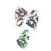
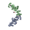
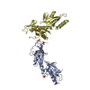
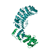
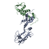
| Remark 300 | BIOMOLECULE THIS ENTRY CONTAINS THE CRYSTALLOGRAPHIC ASYMMETRIC UNIT WHICH CONSISTS OF 4 CHAIN(S). ... BIOMOLECULE THIS ENTRY CONTAINS THE CRYSTALLOGRAPHIC ASYMMETRIC UNIT WHICH CONSISTS OF 4 CHAIN(S). SEE REMARK 350 FOR INFORMATION ON GENERATING THE BIOLOGICAL MOLECULE(S). Each molecule is a part of the SCF complex, which is a hetero tetramer generated by Skp1-Fbs1-Cul1-Rbx1. |
- Structure visualization
| Structure viewer | Molecule: MolmilJmol/JSmol |
|---|
- Downloads & links
-Download
| PDBx/mmCIF format | 2e32.cif.gz | 159.7 KB | Display | PDBx/mmCIF format |
|---|---|---|---|---|
| PDB format | pdb2e32.ent.gz | 125.3 KB | Display | PDB format |
| PDBx/mmJSON format | 2e32.json.gz | Tree view | PDBx/mmJSON format | |
| Others |  Other downloads Other downloads |
-Validation report
| Arichive directory | https://data.pdbj.org/pub/pdb/validation_reports/e3/2e32ftp://data.pdbj.org/pub/pdb/validation_reports/e3/2e32 | HTTPS FTP |
|---|
-Related structure data
| Related structure data |  2e31SC  2e33C S: Starting model for refinement C: citing same article ( |
|---|---|
| Similar structure data |


-Links
 PDBj
PDBj


- Assembly
| Deposited unit | 
| |||||||||||||||||||||||||||||||||||||||||||||||||||||||||||||||||||||||||||||||||||||||||||||||||||||||||||||||||||||||||||||||||||||||||||||||||||||||||||||
|---|---|---|---|---|---|---|---|---|---|---|---|---|---|---|---|---|---|---|---|---|---|---|---|---|---|---|---|---|---|---|---|---|---|---|---|---|---|---|---|---|---|---|---|---|---|---|---|---|---|---|---|---|---|---|---|---|---|---|---|---|---|---|---|---|---|---|---|---|---|---|---|---|---|---|---|---|---|---|---|---|---|---|---|---|---|---|---|---|---|---|---|---|---|---|---|---|---|---|---|---|---|---|---|---|---|---|---|---|---|---|---|---|---|---|---|---|---|---|---|---|---|---|---|---|---|---|---|---|---|---|---|---|---|---|---|---|---|---|---|---|---|---|---|---|---|---|---|---|---|---|---|---|---|---|---|---|---|---|
| 1 | 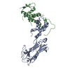
| |||||||||||||||||||||||||||||||||||||||||||||||||||||||||||||||||||||||||||||||||||||||||||||||||||||||||||||||||||||||||||||||||||||||||||||||||||||||||||||
| 2 | 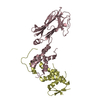
| |||||||||||||||||||||||||||||||||||||||||||||||||||||||||||||||||||||||||||||||||||||||||||||||||||||||||||||||||||||||||||||||||||||||||||||||||||||||||||||
| Unit cell |
| |||||||||||||||||||||||||||||||||||||||||||||||||||||||||||||||||||||||||||||||||||||||||||||||||||||||||||||||||||||||||||||||||||||||||||||||||||||||||||||
| Noncrystallographic symmetry (NCS) | NCS domain:
NCS domain segments: Refine code: 1
NCS ensembles :
|
-Components
| #1: Protein | Mass: 33676.762 Da / Num. of mol.: 2 Source method: isolated from a genetically manipulated source Source: (gene. exp.) Mus musculus (house mouse) / Plasmid: PET28 / Production host:  Escherichia coli (E. coli) / Strain (production host): BL21(RIL) / References: UniProt: Q80UW2 Escherichia coli (E. coli) / Strain (production host): BL21(RIL) / References: UniProt: Q80UW2#2: Protein | Mass: 18972.279 Da / Num. of mol.: 2 Source method: isolated from a genetically manipulated source Source: (gene. exp.) Homo sapiens (human) / Plasmid: PET28 / Production host: Escherichia coli (E. coli) / Strain (production host): BL21(RIL) / References: UniProt: P63208 |
|---|
-Experimental details
-Experiment
| Experiment | Method: X-RAY DIFFRACTION / Number of used crystals: 1 |
|---|
- Sample preparation
| Crystal | Density Matthews: 2.63 Å3/Da / Density % sol: 53.23 % |
|---|---|
| Crystal grow | Temperature: 293 K / Method: vapor diffusion, sitting drop / pH: 5.7 Details: 2.0M ammonium sulphate, 0.1M sodium citrate, 30mM chitobiose, pH 5.7, VAPOR DIFFUSION, SITTING DROP, temperature 293K |
-Data collection
| Diffraction | Mean temperature: 100 K |
|---|---|
| Diffraction source | Source: SYNCHROTRON / Site: SPring-8  / Beamline: BL44XU / Wavelength: 0.9 Å / Beamline: BL44XU / Wavelength: 0.9 Å |
| Detector | Type: Bruker DIP-6040 / Detector: CCD / Date: Jan 29, 2004 |
| Radiation | Protocol: SINGLE WAVELENGTH / Monochromatic (M) / Laue (L): M / Scattering type: x-ray |
| Radiation wavelength | Wavelength: 0.9 Å / Relative weight: 1 |
| Reflection | Resolution: 3.5→152.5 Å / Num. obs: 12279 / % possible obs: 85.2 % / Observed criterion σ(I): 0 / Redundancy: 2.6 % / Biso Wilson estimate: 26.74 Å2 / Rmerge(I) obs: 0.103 / Net I/σ(I): 7 |
| Reflection shell | Resolution: 3.5→3.63 Å / Redundancy: 2.1 % / Rmerge(I) obs: 0.225 / Mean I/σ(I) obs: 4.1 / % possible all: 79 |
- Processing
| Software |
| ||||||||||||||||||||||||||||||||||||||||||||||||||||||||||||||||||||||||||||||||||||||||||||||||||||||||||||||||||||||||||||||||||||||||||||||||||||||||||||||||||||||||||
|---|---|---|---|---|---|---|---|---|---|---|---|---|---|---|---|---|---|---|---|---|---|---|---|---|---|---|---|---|---|---|---|---|---|---|---|---|---|---|---|---|---|---|---|---|---|---|---|---|---|---|---|---|---|---|---|---|---|---|---|---|---|---|---|---|---|---|---|---|---|---|---|---|---|---|---|---|---|---|---|---|---|---|---|---|---|---|---|---|---|---|---|---|---|---|---|---|---|---|---|---|---|---|---|---|---|---|---|---|---|---|---|---|---|---|---|---|---|---|---|---|---|---|---|---|---|---|---|---|---|---|---|---|---|---|---|---|---|---|---|---|---|---|---|---|---|---|---|---|---|---|---|---|---|---|---|---|---|---|---|---|---|---|---|---|---|---|---|---|---|---|---|
| Refinement | Method to determine structure: MOLECULAR REPLACEMENT Starting model: PDB ENTRY 2E31 Resolution: 3.52→56.53 Å / Cor.coef. Fo:Fc: 0.892 / Cor.coef. Fo:Fc free: 0.809 / SU B: 36.134 / SU ML: 0.579 / Cross valid method: THROUGHOUT / ESU R Free: 0.772 / Stereochemistry target values: MAXIMUM LIKELIHOOD / Details: HYDROGENS HAVE BEEN ADDED IN THE RIDING POSITIONS
| ||||||||||||||||||||||||||||||||||||||||||||||||||||||||||||||||||||||||||||||||||||||||||||||||||||||||||||||||||||||||||||||||||||||||||||||||||||||||||||||||||||||||||
| Solvent computation | Ion probe radii: 0.8 Å / Shrinkage radii: 0.8 Å / VDW probe radii: 1.2 Å / Solvent model: MASK | ||||||||||||||||||||||||||||||||||||||||||||||||||||||||||||||||||||||||||||||||||||||||||||||||||||||||||||||||||||||||||||||||||||||||||||||||||||||||||||||||||||||||||
| Displacement parameters | Biso mean: 51.038 Å2
| ||||||||||||||||||||||||||||||||||||||||||||||||||||||||||||||||||||||||||||||||||||||||||||||||||||||||||||||||||||||||||||||||||||||||||||||||||||||||||||||||||||||||||
| Refinement step | Cycle: LAST / Resolution: 3.52→56.53 Å
| ||||||||||||||||||||||||||||||||||||||||||||||||||||||||||||||||||||||||||||||||||||||||||||||||||||||||||||||||||||||||||||||||||||||||||||||||||||||||||||||||||||||||||
| Refine LS restraints |
| ||||||||||||||||||||||||||||||||||||||||||||||||||||||||||||||||||||||||||||||||||||||||||||||||||||||||||||||||||||||||||||||||||||||||||||||||||||||||||||||||||||||||||
| Refine LS restraints NCS | Dom-ID: 1 / Refine-ID: X-RAY DIFFRACTION
| ||||||||||||||||||||||||||||||||||||||||||||||||||||||||||||||||||||||||||||||||||||||||||||||||||||||||||||||||||||||||||||||||||||||||||||||||||||||||||||||||||||||||||
| LS refinement shell | Resolution: 3.516→3.608 Å / Total num. of bins used: 20
|
