 Movie
Movie Controller


+ Open data

- Basic information
| Entry | Database: PDB / ID: 2e1u | ||||||
|---|---|---|---|---|---|---|---|
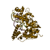
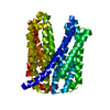
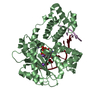
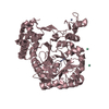
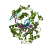
| Title | Crystal structure of Dendranthema morifolium DmAT | ||||||
 Components Components | acyl transferase | ||||||
 Keywords Keywords |  TRANSFERASE / BAHD superfamily / acyl transferase / Dendranthema morifolium / DmAT TRANSFERASE / BAHD superfamily / acyl transferase / Dendranthema morifolium / DmAT | ||||||
| Function / homology | Transferase family / Chloramphenicol Acetyltransferase / Chloramphenicol acetyltransferase-like domain / Chloramphenicol acetyltransferase-like domain superfamily / transferase activity / 2-Layer Sandwich / Alpha Beta / Anthocyanin malonyltransferase homolog Function and homology information Function and homology information | ||||||
| Biological species |  Chrysanthemum x morifolium (florist's chrysanthemum) Chrysanthemum x morifolium (florist's chrysanthemum) | ||||||
| Method | X-RAY DIFFRACTION / SYNCHROTRON / MOLECULAR REPLACEMENT / Resolution: 2.2 Å | ||||||
 Authors Authors | Unno, H. / Ichimaida, F. / Kusunoki, M. / Nakayama, T. | ||||||
 Citation Citation | Journal: J.Biol.Chem. / Year: 2007 Title: Structural and Mutational Studies of Anthocyanin Malonyltransferases Establish the Features of BAHD Enzyme Catalysis Authors: Unno, H. / Ichimaida, F. / Suzuki, H. / Takahashi, S. / Tanaka, Y. / Saito, A. / Nishino, T. / Kusunoki, M. / Nakayama, T. | ||||||
| History |
|
- Structure visualization
| Structure viewer | Molecule: MolmilJmol/JSmol |
|---|
- Downloads & links
-Download
| PDBx/mmCIF format | 2e1u.cif.gz | 195.7 KB | Display | PDBx/mmCIF format |
|---|---|---|---|---|
| PDB format | pdb2e1u.ent.gz | 156.8 KB | Display | PDB format |
| PDBx/mmJSON format | 2e1u.json.gz | Tree view | PDBx/mmJSON format | |
| Others |  Other downloads Other downloads |
-Validation report
| Arichive directory | https://data.pdbj.org/pub/pdb/validation_reports/e1/2e1uftp://data.pdbj.org/pub/pdb/validation_reports/e1/2e1u | HTTPS FTP |
|---|
-Related structure data
| Related structure data | 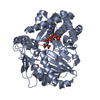 2e1tC 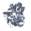 2e1vSC C: citing same article ( S: Starting model for refinement |
|---|---|
| Similar structure data |


-Links
 PDBj
PDBj

- Assembly
| Deposited unit | 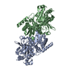
| |||||||||||||||||||||||||||||||||||||||||||||||||||||||||||||||||
|---|---|---|---|---|---|---|---|---|---|---|---|---|---|---|---|---|---|---|---|---|---|---|---|---|---|---|---|---|---|---|---|---|---|---|---|---|---|---|---|---|---|---|---|---|---|---|---|---|---|---|---|---|---|---|---|---|---|---|---|---|---|---|---|---|---|---|
| 1 | 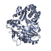
| |||||||||||||||||||||||||||||||||||||||||||||||||||||||||||||||||
| 2 | 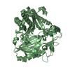
| |||||||||||||||||||||||||||||||||||||||||||||||||||||||||||||||||
| Unit cell |
| |||||||||||||||||||||||||||||||||||||||||||||||||||||||||||||||||
| Noncrystallographic symmetry (NCS) | NCS domain:
NCS domain segments: Ens-ID: 1 / Refine code: 3
|
-Components
| #1: Protein | Mass: 50860.227 Da / Num. of mol.: 2 Source method: isolated from a genetically manipulated source Source: (gene. exp.) Chrysanthemum x morifolium (florist's chrysanthemum)Plasmid: pET15b / Production host:  Escherichia coli (E. coli) / References: UniProt: A4PHY4, Transferases Escherichia coli (E. coli) / References: UniProt: A4PHY4, Transferases#2: Water | ChemComp-HOH / | Water Mass: 18.015 Da / Num. of mol.: 697 / Source method: isolated from a natural source / Formula: H2O Mass: 18.015 Da / Num. of mol.: 697 / Source method: isolated from a natural source / Formula: H2O |
|---|
-Experimental details
-Experiment
| Experiment | Method: X-RAY DIFFRACTION / Number of used crystals: 1 |
|---|
- Sample preparation
| Crystal | Density Matthews: 2.23 Å3/Da / Density % sol: 44.82 % |
|---|---|
| Crystal grow | Temperature: 293 K / Method: vapor diffusion, hanging drop / pH: 6.5 Details: 25% PEG 8000, 10% glycerol, 0.1M ammonium sulfate, 0.1M sodium cacodylate, 10% KCl, pH 6.5, VAPOR DIFFUSION, HANGING DROP, temperature 293K |
-Data collection
| Diffraction | Mean temperature: 90 K |
|---|---|
| Diffraction source | Source: SYNCHROTRON / Site: Photon Factory  / Beamline: BL-5A / Wavelength: 1 Å / Beamline: BL-5A / Wavelength: 1 Å |
| Detector | Type: ADSC QUANTUM 315 / Detector: CCD / Date: Nov 17, 2005 |
| Radiation | Protocol: SINGLE WAVELENGTH / Monochromatic (M) / Laue (L): M / Scattering type: x-ray |
| Radiation wavelength | Wavelength: 1 Å / Relative weight: 1 |
| Reflection | Resolution: 2.2→50 Å / Num. obs: 40795 / % possible obs: 90.2 % / Redundancy: 3.9 % / Biso Wilson estimate: 32.762 Å2 / Rmerge(I) obs: 0.075 / Net I/σ(I): 18.4 |
| Reflection shell | Resolution: 2.2→2.28 Å / Redundancy: 3 % / Rmerge(I) obs: 0.221 / Mean I/σ(I) obs: 3.6 / % possible all: 59.2 |
- Processing
| Software |
| ||||||||||||||||||||||||||||||||||||||||||||||||||||||||||||||||||||||||||||||||||||||||||
|---|---|---|---|---|---|---|---|---|---|---|---|---|---|---|---|---|---|---|---|---|---|---|---|---|---|---|---|---|---|---|---|---|---|---|---|---|---|---|---|---|---|---|---|---|---|---|---|---|---|---|---|---|---|---|---|---|---|---|---|---|---|---|---|---|---|---|---|---|---|---|---|---|---|---|---|---|---|---|---|---|---|---|---|---|---|---|---|---|---|---|---|
| Refinement | Method to determine structure: MOLECULAR REPLACEMENT Starting model: PDB ENTRY 2E1V Resolution: 2.2→33.88 Å / Cor.coef. Fo:Fc: 0.951 / Cor.coef. Fo:Fc free: 0.92 / SU B: 6.37 / SU ML: 0.165 / Cross valid method: THROUGHOUT / ESU R: 0.431 / ESU R Free: 0.255 / Stereochemistry target values: MAXIMUM LIKELIHOOD / Details: HYDROGENS HAVE BEEN ADDED IN THE RIDING POSITIONS
| ||||||||||||||||||||||||||||||||||||||||||||||||||||||||||||||||||||||||||||||||||||||||||
| Solvent computation | Ion probe radii: 0.8 Å / Shrinkage radii: 0.8 Å / VDW probe radii: 1.2 Å / Solvent model: MASK | ||||||||||||||||||||||||||||||||||||||||||||||||||||||||||||||||||||||||||||||||||||||||||
| Displacement parameters | Biso mean: 41.543 Å2
| ||||||||||||||||||||||||||||||||||||||||||||||||||||||||||||||||||||||||||||||||||||||||||
| Refinement step | Cycle: LAST / Resolution: 2.2→33.88 Å
| ||||||||||||||||||||||||||||||||||||||||||||||||||||||||||||||||||||||||||||||||||||||||||
| Refine LS restraints |
| ||||||||||||||||||||||||||||||||||||||||||||||||||||||||||||||||||||||||||||||||||||||||||
| Refine LS restraints NCS | Dom-ID: 1 / Auth asym-ID: A / Ens-ID: 1 / Refine-ID: X-RAY DIFFRACTION
| ||||||||||||||||||||||||||||||||||||||||||||||||||||||||||||||||||||||||||||||||||||||||||
| LS refinement shell | Resolution: 2.2→2.252 Å / Total num. of bins used: 20
|
