 Movie
Movie Controller

[English] 日本語
 Yorodumi
- PDB-2e0c: crystal structure of isocitrate dehydrogenase from Sulfolobus tok... -
+ Open data

- Basic information
| Entry | Database: PDB / ID: 2e0c | ||||||
|---|---|---|---|---|---|---|---|
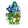
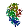
| Title | crystal structure of isocitrate dehydrogenase from Sulfolobus tokodaii strain7 at 2.0 A resolution | ||||||
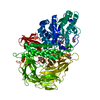
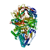
 Components Components | 409aa long hypothetical NADP-dependent isocitrate dehydrogenase | ||||||
 Keywords Keywords |  OXIDOREDUCTASE / homedimer OXIDOREDUCTASE / homedimer | ||||||
| Function / homology |  Function and homology informationisocitrate dehydrogenase (NADP+) / isocitrate dehydrogenase (NADP+) activity / tricarboxylic acid cycle / nucleotide binding / metal ion binding Function and homology informationisocitrate dehydrogenase (NADP+) / isocitrate dehydrogenase (NADP+) activity / tricarboxylic acid cycle / nucleotide binding / metal ion bindingSimilarity search - Function | ||||||
| Biological species |   Sulfolobus tokodaii str. 7 (archaea) Sulfolobus tokodaii str. 7 (archaea) | ||||||
| Method | X-RAY DIFFRACTION / SYNCHROTRON / MOLECULAR REPLACEMENT / Resolution: 2 Å | ||||||
 Authors Authors | Kouyama, T. | ||||||
 Citation Citation | Journal: Archaea / Year: 2018 Title: Crystal Structures of the Putative Isocitrate Dehydrogenase fromSulfolobus tokodaiiStrain 7 in the Apo and NADP+-Bound Forms. Authors: Kondo, H. / Murakami, M. | ||||||
| History |
|
- Structure visualization
| Structure viewer | Molecule: MolmilJmol/JSmol |
|---|
- Downloads & links
-Download
| PDBx/mmCIF format | 2e0c.cif.gz | 175.9 KB | Display | PDBx/mmCIF format |
|---|---|---|---|---|
| PDB format | pdb2e0c.ent.gz | 139.2 KB | Display | PDB format |
| PDBx/mmJSON format | 2e0c.json.gz | Tree view | PDBx/mmJSON format | |
| Others |  Other downloads Other downloads |
-Validation report
| Arichive directory | https://data.pdbj.org/pub/pdb/validation_reports/e0/2e0cftp://data.pdbj.org/pub/pdb/validation_reports/e0/2e0c | HTTPS FTP |
|---|
-Related structure data
| Related structure data | 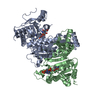 2e5mC  2dhtS S: Starting model for refinement C: citing same article ( |
|---|---|
| Similar structure data |


-Links
 PDBj
PDBj

- Assembly
| Deposited unit | 
| ||||||||
|---|---|---|---|---|---|---|---|---|---|
| 1 |
| ||||||||
| Unit cell |
|
-Components
| #1: Protein | Mass: 46558.871 Da / Num. of mol.: 2 Source method: isolated from a genetically manipulated source Source: (gene. exp.) Sulfolobus tokodaii str. 7 (archaea) / Species: Sulfolobus tokodaii / Strain: strain 7 / Production host:  Escherichia coli (E. coli) Escherichia coli (E. coli)References: UniProt: Q96YK6, isocitrate dehydrogenase (NADP+)#2: Water | ChemComp-HOH / | Water Mass: 18.015 Da / Num. of mol.: 463 / Source method: isolated from a natural source / Formula: H2O Mass: 18.015 Da / Num. of mol.: 463 / Source method: isolated from a natural source / Formula: H2O |
|---|
-Experimental details
-Experiment
| Experiment | Method: X-RAY DIFFRACTION / Number of used crystals: 1 |
|---|
- Sample preparation
| Crystal | Density Matthews: 2.67 Å3/Da / Density % sol: 53.91 % |
|---|---|
| Crystal grow | Temperature: 283 K / Method: vapor diffusion, hanging drop / pH: 7.5 Details: 14% PEG 10000, 0.1M HEPES, pH 7.5, VAPOR DIFFUSION, HANGING DROP, temperature 283K |
-Data collection
| Diffraction | Mean temperature: 100 K |
|---|---|
| Diffraction source | Source: SYNCHROTRON / Site: SPring-8  / Beamline: BL38B1 / Wavelength: 1 Å / Beamline: BL38B1 / Wavelength: 1 Å |
| Detector | Type: ADSC QUANTUM 4 / Detector: CCD / Date: May 14, 2006 |
| Radiation | Monochromator: GRAPHITE / Protocol: SINGLE WAVELENGTH / Monochromatic (M) / Laue (L): M / Scattering type: x-ray |
| Radiation wavelength | Wavelength: 1 Å / Relative weight: 1 |
| Reflection | Resolution: 2→100 Å / Num. all: 66041 / Num. obs: 65823 / % possible obs: 99.5 % / Observed criterion σ(F): 0 / Observed criterion σ(I): 0 / Redundancy: 3.6 % / Biso Wilson estimate: 34.1 Å2 / Rmerge(I) obs: 0.062 / Rsym value: 0.062 / Net I/σ(I): 11.7 |
| Reflection shell | Resolution: 2→2.11 Å / Redundancy: 3.5 % / Rmerge(I) obs: 0.413 / Mean I/σ(I) obs: 2.7 / Num. unique all: 9631 / Rsym value: 0.413 / % possible all: 99.8 |
- Processing
| Software |
| |||||||||||||||||||||||||
|---|---|---|---|---|---|---|---|---|---|---|---|---|---|---|---|---|---|---|---|---|---|---|---|---|---|---|
| Refinement | Method to determine structure: MOLECULAR REPLACEMENT Starting model: 2dht Resolution: 2→15 Å / Isotropic thermal model: Isotropic / Cross valid method: THROUGHOUT / σ(F): 0 / σ(I): 0 / Stereochemistry target values: Engh & Huber
| |||||||||||||||||||||||||
| Displacement parameters | Biso mean: 34.1 Å2
| |||||||||||||||||||||||||
| Refine analyze |
| |||||||||||||||||||||||||
| Refinement step | Cycle: LAST / Resolution: 2→15 Å
| |||||||||||||||||||||||||
| Refine LS restraints |
|
