 Movie
Movie Controller


+ Open data

- Basic information
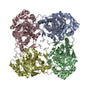
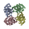
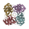
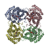
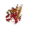
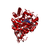
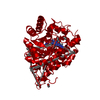
| Entry | Database: PDB / ID: 2du2 | ||||||
|---|---|---|---|---|---|---|---|
| Title | Crystal Structure Analysis of the L-Lactate Oxidase | ||||||
 Components Components | Lactate oxidase | ||||||
 Keywords Keywords |  OXIDOREDUCTASE / TIM BARREL / FMN OXIDOREDUCTASE / TIM BARREL / FMN | ||||||
| Function / homology |  Function and homology informationOxidoreductases; Acting on the CH-OH group of donors; With oxygen as acceptor / lactate oxidation / fatty acid alpha-oxidation / L-lactate dehydrogenase activity / peroxisome / FMN binding / metal ion binding / plasma membrane Function and homology informationOxidoreductases; Acting on the CH-OH group of donors; With oxygen as acceptor / lactate oxidation / fatty acid alpha-oxidation / L-lactate dehydrogenase activity / peroxisome / FMN binding / metal ion binding / plasma membraneSimilarity search - Function | ||||||
| Biological species |  Aerococcus viridans (bacteria) Aerococcus viridans (bacteria) | ||||||
| Method | X-RAY DIFFRACTION / SYNCHROTRON / MOLECULAR REPLACEMENT / Resolution: 2.1 Å | ||||||
 Authors Authors | Morimoto, Y. | ||||||
 Citation Citation | Journal: Biochem.Biophys.Res.Commun. / Year: 2006 Title: The crystal structure of L-lactate oxidase from Aerococcus viridans at 2.1A resolution reveals the mechanism of strict substrate recognition Authors: Umena, Y. / Yorita, K. / Matsuoka, T. / Kita, A. / Fukui, K. / Morimoto, Y. | ||||||
| History |
|
- Structure visualization
| Structure viewer | Molecule: MolmilJmol/JSmol |
|---|
- Downloads & links
-Download
| PDBx/mmCIF format | 2du2.cif.gz | 323.5 KB | Display | PDBx/mmCIF format |
|---|---|---|---|---|
| PDB format | pdb2du2.ent.gz | 257 KB | Display | PDB format |
| PDBx/mmJSON format | 2du2.json.gz | Tree view | PDBx/mmJSON format | |
| Others |  Other downloads Other downloads |
-Validation report
| Arichive directory | https://data.pdbj.org/pub/pdb/validation_reports/du/2du2ftp://data.pdbj.org/pub/pdb/validation_reports/du/2du2 | HTTPS FTP |
|---|
-Related structure data
| Related structure data | 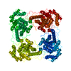 1goxS S: Starting model for refinement |
|---|---|
| Similar structure data |


-Links
 PDBj
PDBj
- Assembly
| Deposited unit | 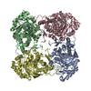
| ||||||||
|---|---|---|---|---|---|---|---|---|---|
| 1 |
| ||||||||
| Unit cell |
|
-Components
| #1: Protein | Mass: 40980.852 Da / Num. of mol.: 4 / Source method: isolated from a natural source / Source: (natural) Aerococcus viridans (bacteria) / References: UniProt: Q44467, lactate 2-monooxygenase#2: Chemical | ChemComp-FMN / Flavin mononucleotide  Mass: 456.344 Da / Num. of mol.: 4 / Source method: obtained synthetically / Formula: C17H21N4O9P Mass: 456.344 Da / Num. of mol.: 4 / Source method: obtained synthetically / Formula: C17H21N4O9P#3: Water | ChemComp-HOH / | Water Mass: 18.015 Da / Num. of mol.: 1109 / Source method: isolated from a natural source / Formula: H2O Mass: 18.015 Da / Num. of mol.: 1109 / Source method: isolated from a natural source / Formula: H2O |
|---|
-Experimental details
-Experiment
| Experiment | Method: X-RAY DIFFRACTION / Number of used crystals: 1 |
|---|
- Sample preparation
| Crystal | Density Matthews: 2.7 Å3/Da / Density % sol: 54.51 % |
|---|---|
| Crystal grow | Temperature: 298 K / Method: vapor diffusion, sitting drop / pH: 8 Details: 18-20%(w/v) PEG 8000, 50mM Tris buffer pH 8.0, VAPOR DIFFUSION, SITTING DROP, temperature 298K |
-Data collection
| Diffraction |
| |||||||||||||||||||||||||
|---|---|---|---|---|---|---|---|---|---|---|---|---|---|---|---|---|---|---|---|---|---|---|---|---|---|---|
| Diffraction source |
| |||||||||||||||||||||||||
| Detector |
| |||||||||||||||||||||||||
| Radiation | Monochromator: Si 111 CHANNEL / Protocol: SINGLE WAVELENGTH / Monochromatic (M) / Laue (L): M / Scattering type: x-ray | |||||||||||||||||||||||||
| Radiation wavelength | Wavelength: 1 Å / Relative weight: 1 | |||||||||||||||||||||||||
| Reflection | Resolution: 2.07→46 Å / Num. all: 108566 / Num. obs: 108566 / % possible obs: 100 % / Observed criterion σ(F): 1 / Observed criterion σ(I): 1 / Redundancy: 12.8 % / Biso Wilson estimate: 19.47 Å2 / Rmerge(I) obs: 0.142 / Net I/σ(I): 22.72 | |||||||||||||||||||||||||
| Reflection shell | Resolution: 2.07→2.14 Å / Rmerge(I) obs: 0.436 / % possible all: 100 |

- Processing
| Software |
| ||||||||||||||||||||||||||||||||||||||||||||||||||||||||||||||||||||||||||||||||||||||||||||||||||||||||||||||||||||||||||||||||||||||||||||||||||||||||||||||||||||||||||
|---|---|---|---|---|---|---|---|---|---|---|---|---|---|---|---|---|---|---|---|---|---|---|---|---|---|---|---|---|---|---|---|---|---|---|---|---|---|---|---|---|---|---|---|---|---|---|---|---|---|---|---|---|---|---|---|---|---|---|---|---|---|---|---|---|---|---|---|---|---|---|---|---|---|---|---|---|---|---|---|---|---|---|---|---|---|---|---|---|---|---|---|---|---|---|---|---|---|---|---|---|---|---|---|---|---|---|---|---|---|---|---|---|---|---|---|---|---|---|---|---|---|---|---|---|---|---|---|---|---|---|---|---|---|---|---|---|---|---|---|---|---|---|---|---|---|---|---|---|---|---|---|---|---|---|---|---|---|---|---|---|---|---|---|---|---|---|---|---|---|---|---|
| Refinement | Method to determine structure: MOLECULAR REPLACEMENT Starting model: PDB ENTRY 1GOX Resolution: 2.1→20 Å / Cor.coef. Fo:Fc: 0.955 / Cor.coef. Fo:Fc free: 0.934 / SU B: 3.384 / SU ML: 0.093 / Isotropic thermal model: Isotropic / Cross valid method: THROUGHOUT / σ(F): 1 / σ(I): 1 / ESU R: 0.167 / ESU R Free: 0.148 / Stereochemistry target values: MAXIMUM LIKELIHOOD
| ||||||||||||||||||||||||||||||||||||||||||||||||||||||||||||||||||||||||||||||||||||||||||||||||||||||||||||||||||||||||||||||||||||||||||||||||||||||||||||||||||||||||||
| Solvent computation | Ion probe radii: 0.8 Å / Shrinkage radii: 0.8 Å / VDW probe radii: 1.2 Å / Solvent model: BABINET MODEL WITH MASK | ||||||||||||||||||||||||||||||||||||||||||||||||||||||||||||||||||||||||||||||||||||||||||||||||||||||||||||||||||||||||||||||||||||||||||||||||||||||||||||||||||||||||||
| Displacement parameters | Biso mean: 19.443 Å2
| ||||||||||||||||||||||||||||||||||||||||||||||||||||||||||||||||||||||||||||||||||||||||||||||||||||||||||||||||||||||||||||||||||||||||||||||||||||||||||||||||||||||||||
| Refinement step | Cycle: LAST / Resolution: 2.1→20 Å
| ||||||||||||||||||||||||||||||||||||||||||||||||||||||||||||||||||||||||||||||||||||||||||||||||||||||||||||||||||||||||||||||||||||||||||||||||||||||||||||||||||||||||||
| Refine LS restraints |
| ||||||||||||||||||||||||||||||||||||||||||||||||||||||||||||||||||||||||||||||||||||||||||||||||||||||||||||||||||||||||||||||||||||||||||||||||||||||||||||||||||||||||||
| LS refinement shell | Resolution: 2.07→2.123 Å / Total num. of bins used: 20
|
