- PDB-2dm1: Solution structure of the second SH3 domain of human protein vav-2 -
+
Open data
ID or keywords:
Loading...
-
Basic information
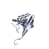
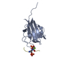
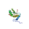
Entry
Database: PDB / ID: 2dm1
Title
Solution structure of the second SH3 domain of human protein vav-2
Components
Protein vav-2
Keywords
SIGNALING PROTEIN / Rho family Guanine nucleotide exchange factor / Structural Genomics / NPPSFA / National Project on Protein Structural and Functional Analyses / RIKEN Structural Genomics/Proteomics Initiative / RSGI
Function / homology
Function and homology information
Azathioprine ADME / regulation of small GTPase mediated signal transduction / lamellipodium assembly / regulation of cell size / epidermal growth factor receptor binding / small GTPase-mediated signal transduction / RHOB GTPase cycle / NRAGE signals death through JNK / regulation of GTPase activity / Fc-gamma receptor signaling pathway involved in phagocytosis ...Azathioprine ADME / regulation of small GTPase mediated signal transduction / lamellipodium assembly / regulation of cell size / epidermal growth factor receptor binding / small GTPase-mediated signal transduction / RHOB GTPase cycle / NRAGE signals death through JNK / regulation of GTPase activity / Fc-gamma receptor signaling pathway involved in phagocytosis / RHOC GTPase cycle / Fc-epsilon receptor signaling pathway / CDC42 GTPase cycle / RHOG GTPase cycle / EPH-ephrin mediated repulsion of cells / RHOA GTPase cycle / RAC2 GTPase cycle / RAC3 GTPase cycle / vascular endothelial growth factor receptor signaling pathway / GPVI-mediated activation cascade / RAC1 GTPase cycle / phosphotyrosine residue binding / FCERI mediated Ca+2 mobilization / guanyl-nucleotide exchange factor activity / VEGFR2 mediated vascular permeability / Signal transduction by L1 / FCERI mediated MAPK activation / FCGR3A-mediated phagocytosis / Regulation of actin dynamics for phagocytic cup formation / platelet activation / VEGFA-VEGFR2 Pathway / G alpha (12/13) signalling events / cell migration / cellular response to xenobiotic stimulus / DAP12 signaling / angiogenesis / positive regulation of phosphatidylinositol 3-kinase/protein kinase B signal transduction / signal transduction / metal ion binding / plasma membrane / cytosol / cytoplasm Similarity search - Function
Conformer selection criteria: target function, structures with the lowest energy, structures with the least restraint violations Conformers calculated total number: 100 / Conformers submitted total number: 20
+
About Yorodumi
-
News
-
Feb 9, 2022. New format data for meta-information of EMDB entries
New format data for meta-information of EMDB entries
Version 3 of the EMDB header file is now the official format.
The previous official version 1.9 will be removed from the archive.
In the structure databanks used in Yorodumi, some data are registered as the other names, "COVID-19 virus" and "2019-nCoV". Here are the details of the virus and the list of structure data.
Jan 31, 2019. EMDB accession codes are about to change! (news from PDBe EMDB page)
EMDB accession codes are about to change! (news from PDBe EMDB page)
The allocation of 4 digits for EMDB accession codes will soon come to an end. Whilst these codes will remain in use, new EMDB accession codes will include an additional digit and will expand incrementally as the available range of codes is exhausted. The current 4-digit format prefixed with “EMD-” (i.e. EMD-XXXX) will advance to a 5-digit format (i.e. EMD-XXXXX), and so on. It is currently estimated that the 4-digit codes will be depleted around Spring 2019, at which point the 5-digit format will come into force.
The EM Navigator/Yorodumi systems omit the EMD- prefix.
Related info.:Q: What is EMD? / ID/Accession-code notation in Yorodumi/EM Navigator
Yorodumi is a browser for structure data from EMDB, PDB, SASBDB, etc.
This page is also the successor to EM Navigator detail page, and also detail information page/front-end page for Omokage search.
The word "yorodu" (or yorozu) is an old Japanese word meaning "ten thousand". "mi" (miru) is to see.
Related info.:EMDB / PDB / SASBDB / Comparison of 3 databanks / Yorodumi Search / Aug 31, 2016. New EM Navigator & Yorodumi / Yorodumi Papers / Jmol/JSmol / Function and homology information / Changes in new EM Navigator and Yorodumi
 Movie
Movie Controller

 Yorodumi
 Open data
 Basic information
 Components
 Keywords
 SIGNALING PROTEIN / Rho family Guanine nucleotide exchange factor /
  Function and homology information
 Authors
 Citation
 Structure visualization
 Downloads & links
 Other downloads
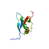

 PDBj
PDBj


 Assembly
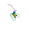
 Sample preparation
 Processing