[English] 日本語
Yorodumi
- PDB-2dln: VANCOMYCIN RESISTANCE: STRUCTURE OF D-ALANINE:D-ALANINE LIGASE AT... -

+
Open data


ID or keywords:

Loading...

-
Basic information

Entry
Database: PDB / ID: 2dln
TitleVANCOMYCIN RESISTANCE: STRUCTURE OF D-ALANINE:D-ALANINE LIGASE AT 2.3 ANGSTROMS RESOLUTION
ComponentsD-ALANINE--D-ALANINE LIGASE
KeywordsLIGASE(PEPTIDOGLYCAN SYNTHESIS)
Function / homology
Function and homology information


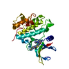
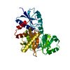
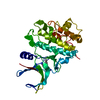
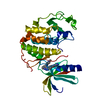
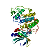
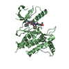
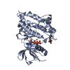
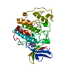
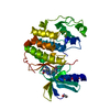
D-alanine-D-alanine ligase / D-alanine-D-alanine ligase activity / peptidoglycan biosynthetic process / cell wall organization / regulation of cell shape / protein homodimerization activity / ATP binding / metal ion binding / cytosol
Similarity search - Function
D-alanine--D-alanine ligase/VANA/B/C, conserved site / D-alanine--D-alanine ligase / D-alanine--D-alanine ligase, C-terminal / D-alanine--D-alanine ligase, N-terminal domain / D-ala D-ala ligase N-terminus / D-ala D-ala ligase C-terminus / D-alanine--D-alanine ligase signature 1. / D-alanine--D-alanine ligase signature 2. / Rossmann fold - #20 / ATP-grasp fold, A domain ...D-alanine--D-alanine ligase/VANA/B/C, conserved site / D-alanine--D-alanine ligase / D-alanine--D-alanine ligase, C-terminal / D-alanine--D-alanine ligase, N-terminal domain / D-ala D-ala ligase N-terminus / D-ala D-ala ligase C-terminus / D-alanine--D-alanine ligase signature 1. / D-alanine--D-alanine ligase signature 2. / Rossmann fold - #20 / ATP-grasp fold, A domain / ATP-grasp fold, B domain / ATP-grasp fold, subdomain 1 / Pre-ATP-grasp domain superfamily / ATP-grasp fold / ATP-grasp fold profile. / D-amino Acid Aminotransferase; Chain A, domain 1 / Dna Ligase; domain 1 / Rossmann fold / 2-Layer Sandwich / 3-Layer(aba) Sandwich / Alpha Beta
Similarity search - Domain/homology
ADENOSINE-5'-DIPHOSPHATE / Chem-PHY / D-alanine--D-alanine ligase B
Similarity search - Component
Biological speciesEscherichia coli (E. coli)
MethodX-RAY DIFFRACTION / Resolution: 2.3 Å
AuthorsKnox, J.R. / Moews, P.C. / Fan, C.
CitationJournal: Science / Year: 1994
Title: Vancomycin resistance: structure of D-alanine:D-alanine ligase at 2.3 A resolution.
Authors: Fan, C. / Moews, P.C. / Walsh, C.T. / Knox, J.R.
History
DepositionJul 18, 1994Processing site: BNL
Revision 1.0Nov 1, 1995Provider: repository / Type: Initial release
Revision 1.1Mar 3, 2008Group: Version format compliance
Revision 1.2Jul 13, 2011Group: Version format compliance
Revision 1.3Jul 17, 2019Group: Data collection / Other / Refinement description / Category: pdbx_database_status / software
Item: _pdbx_database_status.process_site / _software.classification
Revision 1.4Aug 14, 2019Group: Data collection / Refinement description / Category: software / Item: _software.classification
Revision 1.5Feb 14, 2024Group: Data collection / Database references / Derived calculations
Category: chem_comp_atom / chem_comp_bond ...chem_comp_atom / chem_comp_bond / database_2 / pdbx_struct_conn_angle / struct_conn / struct_site
Item: _database_2.pdbx_DOI / _database_2.pdbx_database_accession ..._database_2.pdbx_DOI / _database_2.pdbx_database_accession / _pdbx_struct_conn_angle.ptnr1_auth_comp_id / _pdbx_struct_conn_angle.ptnr1_auth_seq_id / _pdbx_struct_conn_angle.ptnr1_label_asym_id / _pdbx_struct_conn_angle.ptnr1_label_atom_id / _pdbx_struct_conn_angle.ptnr1_label_comp_id / _pdbx_struct_conn_angle.ptnr1_label_seq_id / _pdbx_struct_conn_angle.ptnr3_auth_comp_id / _pdbx_struct_conn_angle.ptnr3_auth_seq_id / _pdbx_struct_conn_angle.ptnr3_label_asym_id / _pdbx_struct_conn_angle.ptnr3_label_atom_id / _pdbx_struct_conn_angle.ptnr3_label_comp_id / _pdbx_struct_conn_angle.ptnr3_label_seq_id / _pdbx_struct_conn_angle.value / _struct_conn.pdbx_dist_value / _struct_conn.ptnr1_auth_comp_id / _struct_conn.ptnr1_auth_seq_id / _struct_conn.ptnr1_label_asym_id / _struct_conn.ptnr1_label_atom_id / _struct_conn.ptnr1_label_comp_id / _struct_conn.ptnr1_label_seq_id / _struct_conn.ptnr2_auth_comp_id / _struct_conn.ptnr2_auth_seq_id / _struct_conn.ptnr2_label_asym_id / _struct_conn.ptnr2_label_atom_id / _struct_conn.ptnr2_label_comp_id / _struct_conn.ptnr2_label_seq_id / _struct_site.pdbx_auth_asym_id / _struct_site.pdbx_auth_comp_id / _struct_site.pdbx_auth_seq_id

-
Structure visualization

Structure viewerMolecule:
MolmilJmol/JSmol

Downloads & links

-
Assembly

Deposited unit
A: D-ALANINE--D-ALANINE LIGASE
hetero molecules


Theoretical massNumber of molelcules
Total (without water)33,6195
Polymers32,8681
Non-polymers7514
Water5,260292
1


  • Idetical with deposited unit
  • defined by author
TypeNameSymmetry operationNumber
identity operation1_555x,y,z1
Unit cell
Length a, b, c (Å)99.300, 51.400, 51.200
Angle α, β, γ (deg.)90.00, 90.00, 90.00
Int Tables number18
Space group name H-MP21212
Atom site foot note1: CIS PROLINE - PRO 140 / 2: CIS PROLINE - PRO 226

-
Components

#1: Protein D-ALANINE--D-ALANINE LIGASE


Mass: 32867.594 Da / Num. of mol.: 1 / Source method: isolated from a natural source / Source: (natural) Escherichia coli (E. coli) / References: UniProt: P07862, D-alanine-D-alanine ligase
#2: Chemical ChemComp-MG / MAGNESIUM ION


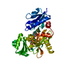
Mass: 24.305 Da / Num. of mol.: 2 / Source method: obtained synthetically / Formula: Mg
#3: Chemical ChemComp-ADP / ADENOSINE-5'-DIPHOSPHATE / Adenosine diphosphate


Mass: 427.201 Da / Num. of mol.: 1 / Source method: obtained synthetically / Formula: C10H15N5O10P2 / Comment: ADP, energy-carrying molecule*YM
#4: Chemical ChemComp-PHY / 1(S)-AMINOETHYL-(2-CARBOXYPROPYL)PHOSPHORYL-PHOSPHINIC ACID


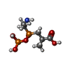
Mass: 275.133 Da / Num. of mol.: 1 / Source method: obtained synthetically / Formula: C6H15NO7P2
#5: Water ChemComp-HOH / water / Water


Mass: 18.015 Da / Num. of mol.: 292 / Source method: isolated from a natural source / Formula: H2O

-
Experimental details

-
Experiment

ExperimentMethod: X-RAY DIFFRACTION

-
Sample preparation

CrystalDensity Matthews: 1.99 Å3/Da / Density % sol: 38.09 %
Crystal grow
*PLUS
pH: 7 / Method: vapor diffusion, hanging drop
Components of the solutions
*PLUS
IDConc.Common nameCrystal-IDSol-IDChemical formula
110 mg/mlenzyme1drop
25 mMATP1drop
35 mM21drop
420 %(v/v)ammonium sulfate1drop
61 mM1dropMgCl2
71 mM1dropNaN3
845 %(v/v)ammonium sulfate1reservoir
5HEPES1drop
9MES1reservoir

-
Data collection

Reflection
*PLUS
Highest resolution: 2.3 Å / Num. obs: 83021 / % possible obs: 90 % / Num. measured all: 11014 / Rmerge(I) obs: 0.057

-
Processing

Software
NameClassification
X-PLORmodel building
PROLSQrefinement
X-PLORrefinement
X-PLORphasing
RefinementResolution: 2.3→15 Å / σ(F): 3
Details: THERE IS NO NON-CRYSTALLOGRAPHIC SYMMETRY. A CLOSE DIMER IS FORMED BY THE TWO-FOLD DIAD OF THE SPACE GROUP (P 21 21 2).
RfactorNum. reflection
Rwork0.172 -
obs0.172 9776
Refinement stepCycle: LAST / Resolution: 2.3→15 Å
ProteinNucleic acidLigandSolventTotal
Num. atoms2305 0 45 292 2642
Refine LS restraints
Refine-IDTypeDev idealDev ideal target
X-RAY DIFFRACTIONx_bond_d0.013
X-RAY DIFFRACTIONx_bond_d_na
X-RAY DIFFRACTIONx_bond_d_prot
X-RAY DIFFRACTIONx_angle_d
X-RAY DIFFRACTIONx_angle_d_na
X-RAY DIFFRACTIONx_angle_d_prot
X-RAY DIFFRACTIONx_angle_deg1.7
X-RAY DIFFRACTIONx_angle_deg_na
X-RAY DIFFRACTIONx_angle_deg_prot
X-RAY DIFFRACTIONx_dihedral_angle_d
X-RAY DIFFRACTIONx_dihedral_angle_d_na
X-RAY DIFFRACTIONx_dihedral_angle_d_prot
X-RAY DIFFRACTIONx_improper_angle_d
X-RAY DIFFRACTIONx_improper_angle_d_na
X-RAY DIFFRACTIONx_improper_angle_d_prot
X-RAY DIFFRACTIONx_mcbond_it0.541
X-RAY DIFFRACTIONx_mcangle_it0.951.5
X-RAY DIFFRACTIONx_scbond_it0.471
X-RAY DIFFRACTIONx_scangle_it0.821.5
Refine LS restraints
*PLUS
Refine-IDTypeDev ideal targetDev ideal
X-RAY DIFFRACTIONx_bond_d0.02
X-RAY DIFFRACTIONx_angle_d0.030.031
X-RAY DIFFRACTIONx_angle_deg
X-RAY DIFFRACTIONx_planar_d0.040.03
X-RAY DIFFRACTIONx_plane_restr0.0180.009
X-RAY DIFFRACTIONx_chiral_restr0.150.149

+
About Yorodumi

-
News

-
Feb 9, 2022. New format data for meta-information of EMDB entries

New format data for meta-information of EMDB entries

  • Version 3 of the EMDB header file is now the official format.
  • The previous official version 1.9 will be removed from the archive.

Related info.:EMDB header

External links:wwPDB to switch to version 3 of the EMDB data model

-
Aug 12, 2020. Covid-19 info

Covid-19 info

URL: https://pdbj.org/emnavi/covid19.php

New page: Covid-19 featured information page in EM Navigator.

Related info.:Covid-19 info / Mar 5, 2020. Novel coronavirus structure data

+
Mar 5, 2020. Novel coronavirus structure data

Novel coronavirus structure data

Related info.:Yorodumi Speices / Aug 12, 2020. Covid-19 info

External links:COVID-19 featured content - PDBj / Molecule of the Month (242):Coronavirus Proteases

+
Jan 31, 2019. EMDB accession codes are about to change! (news from PDBe EMDB page)

EMDB accession codes are about to change! (news from PDBe EMDB page)

  • The allocation of 4 digits for EMDB accession codes will soon come to an end. Whilst these codes will remain in use, new EMDB accession codes will include an additional digit and will expand incrementally as the available range of codes is exhausted. The current 4-digit format prefixed with “EMD-” (i.e. EMD-XXXX) will advance to a 5-digit format (i.e. EMD-XXXXX), and so on. It is currently estimated that the 4-digit codes will be depleted around Spring 2019, at which point the 5-digit format will come into force.
  • The EM Navigator/Yorodumi systems omit the EMD- prefix.

Related info.:Q: What is EMD? / ID/Accession-code notation in Yorodumi/EM Navigator

External links:EMDB Accession Codes are Changing Soon! / Contact to PDBj

+
Jul 12, 2017. Major update of PDB

Major update of PDB

  • wwPDB released updated PDB data conforming to the new PDBx/mmCIF dictionary.
  • This is a major update changing the version number from 4 to 5, and with Remediation, in which all the entries are updated.
  • In this update, many items about electron microscopy experimental information are reorganized (e.g. em_software).
  • Now, EM Navigator and Yorodumi are based on the updated data.

External links:wwPDB Remediation / Enriched Model Files Conforming to OneDep Data Standards Now Available in the PDB FTP Archive

-
Yorodumi

Thousand views of thousand structures

  • Yorodumi is a browser for structure data from EMDB, PDB, SASBDB, etc.
  • This page is also the successor to EM Navigator detail page, and also detail information page/front-end page for Omokage search.
  • The word "yorodu" (or yorozu) is an old Japanese word meaning "ten thousand". "mi" (miru) is to see.

Related info.:EMDB / PDB / SASBDB / Comparison of 3 databanks / Yorodumi Search / Aug 31, 2016. New EM Navigator & Yorodumi / Yorodumi Papers / Jmol/JSmol / Function and homology information / Changes in new EM Navigator and Yorodumi

Read more