 Movie
Movie Controller

[English] 日本語
 Yorodumi
- PDB-2dlb: X-ray Crystal Structure of Protein yopT from Bacillus subtilis. N... -
+ Open data

- Basic information
| Entry | Database: PDB / ID: 2dlb | ||||||
|---|---|---|---|---|---|---|---|
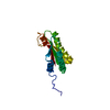
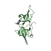
| Title | X-ray Crystal Structure of Protein yopT from Bacillus subtilis. Northeast Structural Genomics Consortium Target SR412 | ||||||
 Components Components | yopT | ||||||
 Keywords Keywords |  STRUCTURAL GENOMICS / UNKNOWN FUNCTION / sr412 / NESG / PSI / Protein Structure Initiative / Northeast Structural Genomics Consortium STRUCTURAL GENOMICS / UNKNOWN FUNCTION / sr412 / NESG / PSI / Protein Structure Initiative / Northeast Structural Genomics Consortium | ||||||
| Function / homology | Protein of unknown function YopT / Protein of unknown function YopT / YopT domain superfamily / Hypothetical protein Yopt / Ubiquitin-like (UB roll) / Roll / Alpha Beta / SPbeta prophage-derived uncharacterized protein YopT Function and homology information Function and homology information | ||||||
| Biological species |  Bacillus subtilis (bacteria) Bacillus subtilis (bacteria) | ||||||
| Method | X-RAY DIFFRACTION / SYNCHROTRON / AB INITIO / Resolution: 1.2 Å | ||||||
 Authors Authors | Kuzin, A.P. / Chen, Y. / Seetharaman, J. / Ho, C.-K. / Cunningham, K. / Janjua, H. / Conover, K. / Ma, L.-C. / Xiao, R. / Acton, T.B. ...Kuzin, A.P. / Chen, Y. / Seetharaman, J. / Ho, C.-K. / Cunningham, K. / Janjua, H. / Conover, K. / Ma, L.-C. / Xiao, R. / Acton, T.B. / Montelione, G.T. / Hunt, J.F. / Tong, L. / Northeast Structural Genomics Consortium (NESG) | ||||||
 Citation Citation | Journal: To be published Title: X-ray structure of hypothetical protein from Bacillus subtilis O34498 at the resolution of 1.2A. NESG target SR412 Authors: Kuzin, A.P. / Chen, Y. / Seetharaman, J. / Ho, C.-K. / Cunningham, K. / Janjua, H. / Conover, K. / Ma, L.-C. / Xiao, R. / Acton, T.B. / Montelione, G.T. / Hunt, J.F. / Tong, L. | ||||||
| History |
|
- Structure visualization
| Structure viewer | Molecule: MolmilJmol/JSmol |
|---|
- Downloads & links
-Download
| PDBx/mmCIF format | 2dlb.cif.gz | 72.5 KB | Display | PDBx/mmCIF format |
|---|---|---|---|---|
| PDB format | pdb2dlb.ent.gz | 57.3 KB | Display | PDB format |
| PDBx/mmJSON format | 2dlb.json.gz | Tree view | PDBx/mmJSON format | |
| Others |  Other downloads Other downloads |
-Validation report
| Arichive directory | https://data.pdbj.org/pub/pdb/validation_reports/dl/2dlbftp://data.pdbj.org/pub/pdb/validation_reports/dl/2dlb | HTTPS FTP |
|---|
-Related structure data
| Similar structure data | |
|---|---|
| Other databases |


-Links
 PDBj
PDBj- Assembly
| Deposited unit | 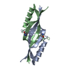
| ||||||||
|---|---|---|---|---|---|---|---|---|---|
| 1 |
| ||||||||
| Unit cell |
| ||||||||
| Details | In according to Dynamic Light Scattering SR412 is dimer. |
-Components
| #1: Protein | Mass: 9249.060 Da / Num. of mol.: 2 Source method: isolated from a genetically manipulated source Source: (gene. exp.) Bacillus subtilis (bacteria) / Plasmid: pE21 / Production host: Escherichia coli (E. coli) / Strain (production host): BL21(DE3)+Magic / References: UniProt: O34498#2: Water | ChemComp-HOH / | Water Mass: 18.015 Da / Num. of mol.: 180 / Source method: isolated from a natural source / Formula: H2O Mass: 18.015 Da / Num. of mol.: 180 / Source method: isolated from a natural source / Formula: H2O |
|---|
-Experimental details
-Experiment
| Experiment | Method: X-RAY DIFFRACTION / Number of used crystals: 1 |
|---|
- Sample preparation
| Crystal | Density Matthews: 2.71 Å3/Da / Density % sol: 52.83 % |
|---|---|
| Crystal grow | Temperature: 277 K / Method: vapor diffusion, hanging drop / pH: 4 Details: 0.1M lithium sulfate, 0.1M sodium citrate, 20% PEG 4000, VAPOR DIFFUSION, HANGING DROP, temperature 277K, pH 4.0 |
-Data collection
| Diffraction | Mean temperature: 100 K | ||||||||||||
|---|---|---|---|---|---|---|---|---|---|---|---|---|---|
| Diffraction source | Source: SYNCHROTRON / Site: NSLS  / Beamline: X4A / Wavelength: 0.9787, 0.9794, 0.9678 / Beamline: X4A / Wavelength: 0.9787, 0.9794, 0.9678 | ||||||||||||
| Detector | Type: ADSC QUANTUM 4 / Detector: CCD / Date: Mar 9, 2006 / Details: mirrors | ||||||||||||
| Radiation | Protocol: MAD / Monochromatic (M) / Laue (L): M / Scattering type: x-ray | ||||||||||||
| Radiation wavelength |
| ||||||||||||
| Reflection | Resolution: 1.2→30 Å / Num. all: 53757 / Num. obs: 53730 / % possible obs: 87.5 % / Observed criterion σ(I): -3 | ||||||||||||
| Reflection shell | Resolution: 1.2→1.24 Å / % possible all: 59.6 |
- Processing
| Software |
| |||||||||||||||||||||||||||||||||
|---|---|---|---|---|---|---|---|---|---|---|---|---|---|---|---|---|---|---|---|---|---|---|---|---|---|---|---|---|---|---|---|---|---|---|
| Refinement | Method to determine structure: AB INITIO / Resolution: 1.2→30 Å / Num. parameters: 11721 / Num. restraintsaints: 14189 / Cross valid method: FREE R / σ(F): 0 / Stereochemistry target values: ENGH AND HUBER Details: Anisotropic refinement reduced free r (no cutoff) by
| |||||||||||||||||||||||||||||||||
| Refine analyze | Num. disordered residues: 6 / Occupancy sum hydrogen: 1100.5 / Occupancy sum non hydrogen: 1281 | |||||||||||||||||||||||||||||||||
| Refinement step | Cycle: LAST / Resolution: 1.2→30 Å
| |||||||||||||||||||||||||||||||||
| Refine LS restraints |
|
