 Movie
Movie Controller

[English] 日本語
 Yorodumi
- PDB-2dkb: DIALKYLGLYCINE DECARBOXYLASE STRUCTURE: BIFUNCTIONAL ACTIVE SITE ... -
+ Open data

- Basic information
| Entry | Database: PDB / ID: 2dkb | ||||||
|---|---|---|---|---|---|---|---|
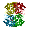
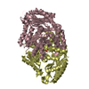
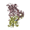
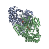
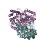
| Title | DIALKYLGLYCINE DECARBOXYLASE STRUCTURE: BIFUNCTIONAL ACTIVE SITE AND ALKALI METAL BINDING SITES | ||||||
 Components Components | 2,2-DIALKYLGLYCINE DECARBOXYLASE (PYRUVATE) 2,2-dialkylglycine decarboxylase (pyruvate) 2,2-dialkylglycine decarboxylase (pyruvate) | ||||||
 Keywords Keywords | LYASE(DECARBOXYLASE) | ||||||
| Function / homology |  Function and homology information2,2-dialkylglycine decarboxylase (pyruvate) / 2,2-dialkylglycine decarboxylase (pyruvate) activity / L-alanine catabolic process, by transamination / alanine-glyoxylate transaminase activity / glyoxylate catabolic process / pyridoxal phosphate binding Function and homology information2,2-dialkylglycine decarboxylase (pyruvate) / 2,2-dialkylglycine decarboxylase (pyruvate) activity / L-alanine catabolic process, by transamination / alanine-glyoxylate transaminase activity / glyoxylate catabolic process / pyridoxal phosphate bindingSimilarity search - Function | ||||||
| Biological species |  Burkholderia cepacia (bacteria) Burkholderia cepacia (bacteria) | ||||||
| Method | X-RAY DIFFRACTION / Resolution: 2.1 Å | ||||||
 Authors Authors | Toney, M.D. / Hohenester, E. / Jansonius, J.N. | ||||||
 Citation Citation | Journal: Science / Year: 1993 Title: Dialkylglycine decarboxylase structure: bifunctional active site and alkali metal sites. Authors: Toney, M.D. / Hohenester, E. / Cowan, S.W. / Jansonius, J.N. #1: Journal: J.Mol.Biol. / Year: 1991Title: Crystallization and Preliminary X-Ray Diffraction Studies of Dialkylglycine Decarboxylase, a Decarboxylating Transaminase Authors: Toney, M.D. / Keller, J.W. / Pauptit, R.A. / Jaeger, J. / Wise, M.K. / Sauder, U. / Jansonius, J.N. #2: Journal: J.Biol.Chem. / Year: 1990Title: Pseudomonas Cepacia 2.2-Dialkylglycine Decarboxylase. Sequence and Expression in Escherichia Coli of Structural and Repressor Genes Authors: Keller, J.W. / Baurick, K.B. / Rutt, G.C. / O'Malley, M.V. / Sonafrank, N.L. / Reynolds, R.A. / Ebbesson, L.O. / Vajdos, F.F. | ||||||
| History |
|
- Structure visualization
| Structure viewer | Molecule: MolmilJmol/JSmol |
|---|
- Downloads & links
-Download
| PDBx/mmCIF format | 2dkb.cif.gz | 96.5 KB | Display | PDBx/mmCIF format |
|---|---|---|---|---|
| PDB format | pdb2dkb.ent.gz | 78.5 KB | Display | PDB format |
| PDBx/mmJSON format | 2dkb.json.gz | Tree view | PDBx/mmJSON format | |
| Others |  Other downloads Other downloads |
-Validation report
| Arichive directory | https://data.pdbj.org/pub/pdb/validation_reports/dk/2dkbftp://data.pdbj.org/pub/pdb/validation_reports/dk/2dkb | HTTPS FTP |
|---|
-Related structure data


-Links
 PDBj
PDBj- Assembly
| Deposited unit | 
| ||||||||
|---|---|---|---|---|---|---|---|---|---|
| 1 | 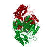
| ||||||||
| 2 | 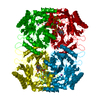
| ||||||||
| Unit cell |
| ||||||||
| Atom site foot note | 1: THE FOLLOWING RESIDUES HAVE WEAK ELECTRON DENSITY: 142 - 143, 346 - 347, AND 369 - 377. 2: AT SITE PLP, RESIDUE LYS 272 HAS THE COFACTOR PYRIDOXAL-5'-PHOSPHATE COVALENTLY ATTACHED TO IT. | ||||||||
| Details | DGD IS A TETRAMER OF IDENTICAL SUBUNITS. THE FOLLOWING TRANSFORMATIONS WILL GENERATE THE OTHER THREE SUBUNITS OF THE TETRAMER: MTRIX1 1 -0.500000 0.866010 0.000000 0.00000 MTRIX2 1 0.866041 0.500000 0.000000 0.00000 MTRIX3 1 0.000000 0.000000 -1.000000 28.86753 MTRIX1 2 0.500000 -0.866010 0.000000 76.34753 MTRIX2 2 -0.866041 -0.500000 0.000000 132.24016 MTRIX3 2 0.000000 0.000000 -1.000000 28.86753 MTRIX1 3 -1.000000 0.000000 0.000000 76.34753 MTRIX2 3 0.000000 -1.000000 0.000000 132.24016 MTRIX3 3 0.000000 0.000000 1.000000 0.00000 |
-Components
| #1: Protein | 2,2-dialkylglycine decarboxylase (pyruvate) Mass: 46577.402 Da / Num. of mol.: 1 Source method: isolated from a genetically manipulated source Source: (gene. exp.) Burkholderia cepacia (bacteria) / Production host: Escherichia coli (E. coli)References: UniProt: P16932, 2,2-dialkylglycine decarboxylase (pyruvate) | ||||||||
|---|---|---|---|---|---|---|---|---|---|
| #2: Chemical |   Mass: 22.990 Da / Num. of mol.: 2 / Source method: obtained synthetically / Formula: Na Mass: 22.990 Da / Num. of mol.: 2 / Source method: obtained synthetically / Formula: Na#3: Chemical | ChemComp-PLP / | Pyridoxal phosphate  Mass: 247.142 Da / Num. of mol.: 1 / Source method: obtained synthetically / Formula: C8H10NO6P Mass: 247.142 Da / Num. of mol.: 1 / Source method: obtained synthetically / Formula: C8H10NO6P#4: Chemical | ChemComp-MES / | MES (buffer)  Mass: 195.237 Da / Num. of mol.: 1 / Source method: obtained synthetically / Formula: C6H13NO4S / Comment: pH buffer*YM Mass: 195.237 Da / Num. of mol.: 1 / Source method: obtained synthetically / Formula: C6H13NO4S / Comment: pH buffer*YM#5: Water | ChemComp-HOH / | Water Mass: 18.015 Da / Num. of mol.: 229 / Source method: isolated from a natural source / Formula: H2O Mass: 18.015 Da / Num. of mol.: 229 / Source method: isolated from a natural source / Formula: H2OSequence details | SEQUENCE ADVISORY NOTICE DIFFERENCE BETWEEN SWISS-PROT AND PDB SEQUENCE. SWISS-PROT ENTRY NAME: ...SEQUENCE ADVISORY NOTICE DIFFERENCE | |
-Experimental details
-Experiment
| Experiment | Method: X-RAY DIFFRACTION |
|---|
- Sample preparation
| Crystal | Density Matthews: 3.13 Å3/Da / Density % sol: 60.67 % | ||||||||||||||||||||||||||||||||||||||||||||||||||||||||||||||||||
|---|---|---|---|---|---|---|---|---|---|---|---|---|---|---|---|---|---|---|---|---|---|---|---|---|---|---|---|---|---|---|---|---|---|---|---|---|---|---|---|---|---|---|---|---|---|---|---|---|---|---|---|---|---|---|---|---|---|---|---|---|---|---|---|---|---|---|---|
| Crystal | *PLUS Density % sol: 61 % | ||||||||||||||||||||||||||||||||||||||||||||||||||||||||||||||||||
| Crystal grow | *PLUS pH: 7.5 / Method: vapor diffusion, hanging dropDetails: taken from Toney, M.D., (1991) J.Mol.Biol., 222, 873. | ||||||||||||||||||||||||||||||||||||||||||||||||||||||||||||||||||
| Components of the solutions | *PLUS
|
-Data collection
| Reflection | *PLUS Highest resolution: 2.1 Å / Num. obs: 32665 / % possible obs: 93 % / Rmerge(I) obs: 0.078 |
|---|
- Processing
| Software | Name: TNT / Classification: refinement | ||||||||||||||||||||||||||||||
|---|---|---|---|---|---|---|---|---|---|---|---|---|---|---|---|---|---|---|---|---|---|---|---|---|---|---|---|---|---|---|---|
| Refinement | Resolution: 2.1→8 Å / Rfactor obs: 0.178 Details: ATOMS THAT ARE NOT WELL DEFINED DUE TO POOR ELECTRON DENSITY OR REFINE TO B-FACTORS (GREATER THAN) 100 ANGSTROMS**2 HAVE A WEIGHT OF ZERO. | ||||||||||||||||||||||||||||||
| Refinement step | Cycle: LAST / Resolution: 2.1→8 Å
| ||||||||||||||||||||||||||||||
| Refine LS restraints |
| ||||||||||||||||||||||||||||||
| Software | *PLUS Name: TNT / Classification: refinement | ||||||||||||||||||||||||||||||
| Refinement | *PLUS Rfactor obs: 0.178 | ||||||||||||||||||||||||||||||
| Solvent computation | *PLUS | ||||||||||||||||||||||||||||||
| Displacement parameters | *PLUS | ||||||||||||||||||||||||||||||
| Refine LS restraints | *PLUS
|
