 Movie
Movie Controller

[English] 日本語
 Yorodumi
- PDB-2dhs: Solution Structure of Nucleic Acid Binding Protein CUGBP1ab and i... -
+ Open data

- Basic information
| Entry | Database: PDB / ID: 2dhs | ||||||
|---|---|---|---|---|---|---|---|
| Title | Solution Structure of Nucleic Acid Binding Protein CUGBP1ab and its Binding Study with DNA and RNA | ||||||
 Components Components | CUG triplet repeat RNA-binding protein 1 | ||||||
 Keywords Keywords |  RNA BINDING PROTEIN / beta sheet / two helices packed against the beta sheet RNA BINDING PROTEIN / beta sheet / two helices packed against the beta sheet | ||||||
| Function / homology |  Function and homology information Function and homology informationBRE binding / perinucleolar compartment / post-transcriptional gene silencing / regulatory ncRNA-mediated post-transcriptional gene silencing / mRNA splice site recognition / pre-mRNA binding / embryo development ending in birth or egg hatching / mRNA destabilization / regulation of alternative mRNA splicing, via spliceosome / germ cell development ...BRE binding / perinucleolar compartment / post-transcriptional gene silencing / regulatory ncRNA-mediated post-transcriptional gene silencing / mRNA splice site recognition / pre-mRNA binding / embryo development ending in birth or egg hatching / mRNA destabilization / regulation of alternative mRNA splicing, via spliceosome / germ cell development / regulation of RNA splicing / mRNA regulatory element binding translation repressor activity / mRNA 3'-UTR binding / mRNA processing / cytoplasmic stress granule / regulation of inflammatory response / ribonucleoprotein complex / negative regulation of cell population proliferation / negative regulation of gene expression / mRNA binding / positive regulation of gene expression / RNA binding / nucleoplasm / membrane / nucleus / cytoplasmSimilarity search - Function | ||||||
| Biological species |  Homo sapiens (human) Homo sapiens (human) | ||||||
| Method | SOLUTION NMR / simulated annealing, molecular dynamics | ||||||
 Authors Authors | Xia, Y.L. / Jun, K.Y. / Zhu, Q. / Han, X.G. / Zhang, H. / Timchenko, L. / Swanson, M. / Gao, X.L. | ||||||
 Citation Citation | Journal: To be Published Title: Solution Structure of Nucleic Acid Binding Protein CUGBP1ab and its Binding Study with DNA and RNA Authors: Xia, Y.L. / Jun, K.Y. / Zhu, Q. / Han, X.G. / Zhang, H. / Timchenko, L. / Swanson, M. / Gao, X.L. | ||||||
| History |
|
- Structure visualization
| Structure viewer | Molecule: MolmilJmol/JSmol |
|---|
- Downloads & links
-Download
| PDBx/mmCIF format | 2dhs.cif.gz | 457.4 KB | Display | PDBx/mmCIF format |
|---|---|---|---|---|
| PDB format | pdb2dhs.ent.gz | 387.8 KB | Display | PDB format |
| PDBx/mmJSON format | 2dhs.json.gz | Tree view | PDBx/mmJSON format | |
| Others |  Other downloads Other downloads |
-Validation report
| Arichive directory | https://data.pdbj.org/pub/pdb/validation_reports/dh/2dhsftp://data.pdbj.org/pub/pdb/validation_reports/dh/2dhs | HTTPS FTP |
|---|
-Related structure data
| Similar structure data | |
|---|---|
| Other databases |
|


-Links
 PDBj
PDBj- Assembly
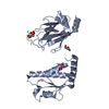
| Deposited unit | 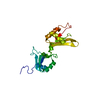
| |||||||||
|---|---|---|---|---|---|---|---|---|---|---|
| 1 |
| |||||||||
| NMR ensembles |
|
-Components
| #1: Protein | Mass: 21106.293 Da / Num. of mol.: 1 / Fragment: RRM1 and RRM2 Source method: isolated from a genetically manipulated source Source: (gene. exp.) Homo sapiens (human) / Gene: CUGBP1 (AMINO ACIDS 1 - 187) / Plasmid: pET15b / Species (production host): Escherichia coli / Production host:  Escherichia coli BL21(DE3) (bacteria) / Strain (production host): BL21 (DE3) / References: UniProt: Q92879 Escherichia coli BL21(DE3) (bacteria) / Strain (production host): BL21 (DE3) / References: UniProt: Q92879 |
|---|
-Experimental details
-Experiment
| Experiment | Method: SOLUTION NMR | ||||||||||||||||||||||||||||||||||||||||||||||||||||||||||||
|---|---|---|---|---|---|---|---|---|---|---|---|---|---|---|---|---|---|---|---|---|---|---|---|---|---|---|---|---|---|---|---|---|---|---|---|---|---|---|---|---|---|---|---|---|---|---|---|---|---|---|---|---|---|---|---|---|---|---|---|---|---|
| NMR experiment |
| ||||||||||||||||||||||||||||||||||||||||||||||||||||||||||||
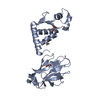
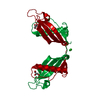
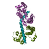
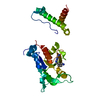
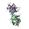
| NMR details | Text: The structure was determined using triple-resonance NMR spectroscopy. The both two domains RRM1 (RNA recognition motif) and RRM2 contain the same beta-alpha-beta-beta-alpha-beta topology as ...Text: The structure was determined using triple-resonance NMR spectroscopy. The both two domains RRM1 (RNA recognition motif) and RRM2 contain the same beta-alpha-beta-beta-alpha-beta topology as expected for RNA-recognition motif. The four beta strands form an anti parallel beta sheet. The two alpha helices are packed against the beta sheet. Other NMR experiments including 15N T1 and T2 and {1H}-15N steady state NOE, and titration with DNA and RNA, were carried out. the RRM1 shows more conformat on exchange than RRM2. The N-terminal region and the linker between the RRM1 and RRM2 show more flexibility than other regions. In addition, two loops L3 and L5 in RRM1 also demonstrate considerable flexibility and conformation exchange. The conformation exchange and flexibility in the RRM1 may be utilized in the recognition process by allowing different conformational states to be accessed and facilitating induced fit for complex forming. From the titration experiments,we found that the (GTC)3 and (GUCU)3 mainly bind the two central beta strands of the RRM1 and the linker of the RRM1 and RRM2 domains. |
- Sample preparation
| Details |
| |||||||||
|---|---|---|---|---|---|---|---|---|---|---|
| Sample conditions | Ionic strength: 25mM sodium phosphate, 50mM NaCl, 0.25mM NaN3, 0.125mM EDTA pH: 5.8 / Pressure: ambient / Temperature: 293 K |
-NMR measurement
| NMR spectrometer |
|
|---|
- Processing
| NMR software |
| ||||||||||||||||||||
|---|---|---|---|---|---|---|---|---|---|---|---|---|---|---|---|---|---|---|---|---|---|
| Refinement | Method: simulated annealing, molecular dynamics / Software ordinal: 1 Details: The protein CUGBP1ab has total 187 residues (residues 1-187 of CUGBP1) including an N-terminus Histag-thrombin site (21 amino acids) and 20 amino acids in the C-terminus which is from the ...Details: The protein CUGBP1ab has total 187 residues (residues 1-187 of CUGBP1) including an N-terminus Histag-thrombin site (21 amino acids) and 20 amino acids in the C-terminus which is from the plasmid. The molecular weight of the CUGBP1ab is 25.4 kDa. The structures are based on a total of 4228 restraints, 3776 are NOE-derived distance donstraints, 324 dihedral angle restraints,128 distance restraints from hydrogen bonds. | ||||||||||||||||||||
| NMR representative | Selection criteria: lowest energy | ||||||||||||||||||||
| NMR ensemble | Conformer selection criteria: structures with the lowest energy Conformers calculated total number: 20 / Conformers submitted total number: 7 |
