 Movie
Movie Controller

[English] 日本語
 Yorodumi
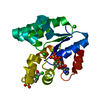
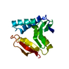
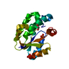
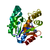
- PDB-2dfn: Structure of shikimate kinase from Mycobacterium tuberculosis com... -
+ Open data

- Basic information
| Entry | Database: PDB / ID: 2dfn | ||||||
|---|---|---|---|---|---|---|---|
| Title | Structure of shikimate kinase from Mycobacterium tuberculosis complexed with ADP and shikimate at 1.9 angstrons of resolution | ||||||
 Components Components | Shikimate kinase | ||||||
 Keywords Keywords | TRANSFERASE / alpha/beta / shikimate pathway | ||||||
| Function / homology |  Function and homology informationshikimate kinase / shikimate metabolic process / Chorismate via Shikimate Pathway / shikimate kinase activity / chorismate biosynthetic process / aromatic amino acid family biosynthetic process / amino acid biosynthetic process / phosphorylation / magnesium ion binding / ATP binding / cytosol Function and homology informationshikimate kinase / shikimate metabolic process / Chorismate via Shikimate Pathway / shikimate kinase activity / chorismate biosynthetic process / aromatic amino acid family biosynthetic process / amino acid biosynthetic process / phosphorylation / magnesium ion binding / ATP binding / cytosolSimilarity search - Function | ||||||
| Biological species |   Mycobacterium tuberculosis (bacteria) Mycobacterium tuberculosis (bacteria) | ||||||
| Method | X-RAY DIFFRACTION / SYNCHROTRON / MOLECULAR REPLACEMENT / Resolution: 1.93 Å | ||||||
 Authors Authors | Dias, M.V. / Faim, L.M. / Vasconcelos, I.B. / de Oliveira, J.S. / Basso, L.A. / Santos, D.S. / de Azevedo, W.F. | ||||||
 Citation Citation | Journal: ACTA CRYSTALLOGR.,SECT.F / Year: 2007 Title: Effects of the magnesium and chloride ions and shikimate on the structure of shikimate kinase from Mycobacterium tuberculosis Authors: Dias, M.V. / Faim, L.M. / Vasconcelos, I.B. / de Oliveira, J.S. / Basso, L.A. / Santos, D.S. / de Azevedo, W.F. | ||||||
| History |
|
- Structure visualization
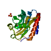
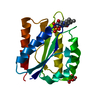
| Structure viewer | Molecule: MolmilJmol/JSmol |
|---|
- Downloads & links
-Download
| PDBx/mmCIF format | 2dfn.cif.gz | 51.7 KB | Display | PDBx/mmCIF format |
|---|---|---|---|---|
| PDB format | pdb2dfn.ent.gz | 36 KB | Display | PDB format |
| PDBx/mmJSON format | 2dfn.json.gz | Tree view | PDBx/mmJSON format | |
| Others |  Other downloads Other downloads |
-Validation report
| Arichive directory | https://data.pdbj.org/pub/pdb/validation_reports/df/2dfnftp://data.pdbj.org/pub/pdb/validation_reports/df/2dfn | HTTPS FTP |
|---|
-Related structure data
| Related structure data |  2dftC 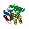 1we2S C: citing same article ( S: Starting model for refinement |
|---|---|
| Similar structure data |


-Links
 PDBj
PDBj
- Assembly
| Deposited unit | 
| ||||||||
|---|---|---|---|---|---|---|---|---|---|
| 1 |
| ||||||||
| Unit cell |
|
-Components
| #1: Protein | / SK Mass: 18612.352 Da / Num. of mol.: 1 Source method: isolated from a genetically manipulated source Source: (gene. exp.) Mycobacterium tuberculosis (bacteria) / Gene: AroK / Production host: Escherichia coli (E. coli)References: UniProt: P0A4Z2, UniProt: P9WPY3*PLUS, shikimate kinase |
|---|---|
| #2: Chemical | ChemComp-CL / Chloride  Mass: 35.453 Da / Num. of mol.: 1 / Source method: obtained synthetically / Formula: Cl Mass: 35.453 Da / Num. of mol.: 1 / Source method: obtained synthetically / Formula: Cl |
| #3: Chemical | ChemComp-ADP / Adenosine diphosphate  Mass: 427.201 Da / Num. of mol.: 1 / Source method: obtained synthetically / Formula: C10H15N5O10P2 / Comment: ADP, energy-carrying molecule*YM Mass: 427.201 Da / Num. of mol.: 1 / Source method: obtained synthetically / Formula: C10H15N5O10P2 / Comment: ADP, energy-carrying molecule*YM |
| #4: Chemical | ChemComp-SKM / (Shikimic acid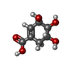  Mass: 174.151 Da / Num. of mol.: 1 / Source method: obtained synthetically / Formula: C7H10O5 Mass: 174.151 Da / Num. of mol.: 1 / Source method: obtained synthetically / Formula: C7H10O5 |
| #5: Water | ChemComp-HOH / Water Mass: 18.015 Da / Num. of mol.: 192 / Source method: isolated from a natural source / Formula: H2O Mass: 18.015 Da / Num. of mol.: 192 / Source method: isolated from a natural source / Formula: H2O |
-Experimental details
-Experiment
| Experiment | Method: X-RAY DIFFRACTION / Number of used crystals: 1 |
|---|
- Sample preparation
| Crystal | Density Matthews: 2.84 Å3/Da / Density % sol: 56.76 % |
|---|---|
| Crystal grow | Temperature: 293 K / Method: vapor diffusion / pH: 8 Details: 0.1M tris-HCl, 17% PEG 1500, 0.5-0.7M LiCl, pH 8.0, VAPOR DIFFUSION, temperature 293K |
-Data collection
| Diffraction | Mean temperature: 100 K |
|---|---|
| Diffraction source | Source: SYNCHROTRON / Site: LNLS  / Beamline: D03B-MX1 / Wavelength: 1.427 Å / Beamline: D03B-MX1 / Wavelength: 1.427 Å |
| Detector | Type: RIGAKU / Detector: IMAGE PLATE / Date: Jun 7, 2005 |
| Radiation | Protocol: SINGLE WAVELENGTH / Monochromatic (M) / Laue (L): M / Scattering type: x-ray |
| Radiation wavelength | Wavelength: 1.427 Å / Relative weight: 1 |
| Reflection | Resolution: 1.93→35.16 Å / Num. all: 16169 / Num. obs: 16017 / % possible obs: 99 % / Observed criterion σ(F): 2 / Observed criterion σ(I): 2 / Net I/σ(I): 4.8 |
| Reflection shell | Resolution: 1.93→2.03 Å / Redundancy: 4.6 % / Num. unique all: 2160 / % possible all: 21.6 |
- Processing
| Software |
| ||||||||||||||||||||||||||||||||||||||||||||||||||||||||||||||||||||||||||||||||||||||||||
|---|---|---|---|---|---|---|---|---|---|---|---|---|---|---|---|---|---|---|---|---|---|---|---|---|---|---|---|---|---|---|---|---|---|---|---|---|---|---|---|---|---|---|---|---|---|---|---|---|---|---|---|---|---|---|---|---|---|---|---|---|---|---|---|---|---|---|---|---|---|---|---|---|---|---|---|---|---|---|---|---|---|---|---|---|---|---|---|---|---|---|---|
| Refinement | Method to determine structure: MOLECULAR REPLACEMENT Starting model: PDB ENTRY 1WE2 Resolution: 1.93→35.16 Å / Cor.coef. Fo:Fc: 0.951 / Cor.coef. Fo:Fc free: 0.914 / SU B: 3.963 / SU ML: 0.116 / Cross valid method: THROUGHOUT / σ(F): 2 / ESU R: 0.159 / ESU R Free: 0.165 / Stereochemistry target values: MAXIMUM LIKELIHOOD / Details: HYDROGENS HAVE BEEN ADDED IN THE RIDING POSITIONS
| ||||||||||||||||||||||||||||||||||||||||||||||||||||||||||||||||||||||||||||||||||||||||||
| Solvent computation | Ion probe radii: 0.8 Å / Shrinkage radii: 0.8 Å / VDW probe radii: 1.2 Å / Solvent model: MASK | ||||||||||||||||||||||||||||||||||||||||||||||||||||||||||||||||||||||||||||||||||||||||||
| Displacement parameters | Biso mean: 32.718 Å2
| ||||||||||||||||||||||||||||||||||||||||||||||||||||||||||||||||||||||||||||||||||||||||||
| Refinement step | Cycle: LAST / Resolution: 1.93→35.16 Å
| ||||||||||||||||||||||||||||||||||||||||||||||||||||||||||||||||||||||||||||||||||||||||||
| Refine LS restraints |
| ||||||||||||||||||||||||||||||||||||||||||||||||||||||||||||||||||||||||||||||||||||||||||
| LS refinement shell | Resolution: 1.93→1.98 Å / Total num. of bins used: 20
|
