 Movie
Movie Controller

[English] 日本語
 Yorodumi
- PDB-2dds: Crystal structure of sphingomyelinase from Bacillus cereus with c... -
+ Open data

- Basic information
| Entry | Database: PDB / ID: 2dds | ||||||
|---|---|---|---|---|---|---|---|
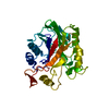
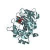
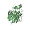
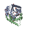
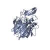
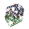
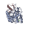
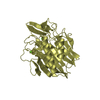
| Title | Crystal structure of sphingomyelinase from Bacillus cereus with cobalt ion | ||||||
 Components Components | Sphingomyelin phosphodiesterase | ||||||
 Keywords Keywords | HYDROLASE / DNase I like folding / RIKEN Structural Genomics/Proteomics Initiative / RSGI / Structural Genomics | ||||||
| Function / homology |  Function and homology informationsphingomyelin phosphodiesterase / sphingomyelin phosphodiesterase activity / killing of cells of another organism / extracellular region Function and homology informationsphingomyelin phosphodiesterase / sphingomyelin phosphodiesterase activity / killing of cells of another organism / extracellular regionSimilarity search - Function | ||||||
| Biological species |  Bacillus cereus (bacteria) Bacillus cereus (bacteria) | ||||||
| Method | X-RAY DIFFRACTION / MOLECULAR REPLACEMENT / Resolution: 1.8 Å | ||||||
 Authors Authors | Ago, H. / Oda, M. / Takahashi, M. / Tsuge, H. / Ochi, S. / Katunuma, N. / Miyano, M. / Sakurai, J. / RIKEN Structural Genomics/Proteomics Initiative (RSGI) | ||||||
 Citation Citation | Journal: J.Biol.Chem. / Year: 2006 Title: Structural Basis of the Sphingomyelin Phosphodiesterase Activity in Neutral Sphingomyelinase from Bacillus cereus. Authors: Ago, H. / Oda, M. / Takahashi, M. / Tsuge, H. / Ochi, S. / Katunuma, N. / Miyano, M. / Sakurai, J. | ||||||
| History |
|
- Structure visualization
| Structure viewer | Molecule: MolmilJmol/JSmol |
|---|
- Downloads & links
-Download
| PDBx/mmCIF format | 2dds.cif.gz | 260.6 KB | Display | PDBx/mmCIF format |
|---|---|---|---|---|
| PDB format | pdb2dds.ent.gz | 215.2 KB | Display | PDB format |
| PDBx/mmJSON format | 2dds.json.gz | Tree view | PDBx/mmJSON format | |
| Others |  Other downloads Other downloads |
-Validation report
| Arichive directory | https://data.pdbj.org/pub/pdb/validation_reports/dd/2ddsftp://data.pdbj.org/pub/pdb/validation_reports/dd/2dds | HTTPS FTP |
|---|
-Related structure data


-Links
 PDBj
PDBj


- Assembly
| Deposited unit | 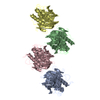
| ||||||||
|---|---|---|---|---|---|---|---|---|---|
| 1 | 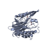
| ||||||||
| 2 | 
| ||||||||
| 3 | 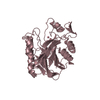
| ||||||||
| 4 | 
| ||||||||
| Unit cell |
|
-Components
| #1: Protein | / sphingomyelinase Mass: 34343.004 Da / Num. of mol.: 4 Source method: isolated from a genetically manipulated source Source: (gene. exp.) Bacillus cereus (bacteria) / Strain: IAM 1029 / Plasmid: pHY300PLK / Production host: Bacillus subtilis (bacteria) / Strain (production host): ISW1214References: UniProt: P11889, sphingomyelin phosphodiesterase#2: Chemical | ChemComp-CO /   Mass: 58.933 Da / Num. of mol.: 12 / Source method: obtained synthetically / Formula: Co Mass: 58.933 Da / Num. of mol.: 12 / Source method: obtained synthetically / Formula: Co#3: Water | ChemComp-HOH / | Water Mass: 18.015 Da / Num. of mol.: 1116 / Source method: isolated from a natural source / Formula: H2O Mass: 18.015 Da / Num. of mol.: 1116 / Source method: isolated from a natural source / Formula: H2O |
|---|
-Experimental details
-Experiment
| Experiment | Method: X-RAY DIFFRACTION / Number of used crystals: 1 |
|---|
- Sample preparation
| Crystal | Density Matthews: 2.18 Å3/Da / Density % sol: 43.61 % |
|---|---|
| Crystal grow | Temperature: 283 K / Method: vapor diffusion / pH: 6.5 Details: 18% (w/v) PEG 8000, 0.2M cobalt chloride, 0.1M MES, pH 6.5, VAPOR DIFFUSION, temperature 283K |
-Data collection
| Diffraction | Mean temperature: 100 K |
|---|---|
| Diffraction source | Source: ROTATING ANODE / Type: RIGAKU / Wavelength: 1.5418 Å |
| Detector | Type: RIGAKU RAXIS VII / Detector: IMAGE PLATE / Date: Aug 14, 2004 |
| Radiation | Protocol: SINGLE WAVELENGTH / Monochromatic (M) / Laue (L): M / Scattering type: x-ray |
| Radiation wavelength | Wavelength: 1.5418 Å / Relative weight: 1 |
| Reflection | Resolution: 1.77→35.8 Å / Num. all: 103230 / Num. obs: 103230 / % possible obs: 91.1 % / Biso Wilson estimate: 15.8 Å2 |
| Reflection shell | Resolution: 1.77→1.83 Å / % possible all: 83.6 |
- Processing
| Software |
| ||||||||||||||||||||||||||||||||||||||||||||||||||||||||||||||||||||||||||||||||
|---|---|---|---|---|---|---|---|---|---|---|---|---|---|---|---|---|---|---|---|---|---|---|---|---|---|---|---|---|---|---|---|---|---|---|---|---|---|---|---|---|---|---|---|---|---|---|---|---|---|---|---|---|---|---|---|---|---|---|---|---|---|---|---|---|---|---|---|---|---|---|---|---|---|---|---|---|---|---|---|---|---|
| Refinement | Method to determine structure: MOLECULAR REPLACEMENT / Resolution: 1.8→19.67 Å / Rfactor Rfree error: 0.002 / Data cutoff high absF: 1415548.83 / Data cutoff low absF: 0 / Isotropic thermal model: RESTRAINED / Cross valid method: THROUGHOUT / σ(F): 0
| ||||||||||||||||||||||||||||||||||||||||||||||||||||||||||||||||||||||||||||||||
| Solvent computation | Solvent model: FLAT MODEL / Bsol: 32.0242 Å2 / ksol: 0.324019 e/Å3 | ||||||||||||||||||||||||||||||||||||||||||||||||||||||||||||||||||||||||||||||||
| Displacement parameters | Biso mean: 18.7 Å2
| ||||||||||||||||||||||||||||||||||||||||||||||||||||||||||||||||||||||||||||||||
| Refine analyze |
| ||||||||||||||||||||||||||||||||||||||||||||||||||||||||||||||||||||||||||||||||
| Refinement step | Cycle: LAST / Resolution: 1.8→19.67 Å
| ||||||||||||||||||||||||||||||||||||||||||||||||||||||||||||||||||||||||||||||||
| Refine LS restraints |
| ||||||||||||||||||||||||||||||||||||||||||||||||||||||||||||||||||||||||||||||||
| LS refinement shell | Resolution: 1.8→1.91 Å / Rfactor Rfree error: 0.008 / Total num. of bins used: 6
| ||||||||||||||||||||||||||||||||||||||||||||||||||||||||||||||||||||||||||||||||
| Xplor file |
|
