 Movie
Movie Controller

[English] 日本語
 Yorodumi
- PDB-2d60: Crystal structure of deoxy human hemoglobin complexed with two L3... -
+ Open data

- Basic information
| Entry | Database: PDB / ID: 2d60 | ||||||
|---|---|---|---|---|---|---|---|
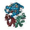
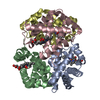
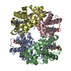
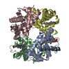
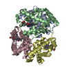
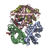
| Title | Crystal structure of deoxy human hemoglobin complexed with two L35 molecules | ||||||
 Components Components |
| ||||||
 Keywords Keywords | OXYGEN STORAGE/TRANSPORT /  HEMOGLOBIN / L35 / ALLOSTERIC EFFECTOR / CRYSTAL SRUCTURE / OXYGEN STORAGE-TRANSPORT COMPLEX HEMOGLOBIN / L35 / ALLOSTERIC EFFECTOR / CRYSTAL SRUCTURE / OXYGEN STORAGE-TRANSPORT COMPLEX | ||||||
| Function / homology |  Function and homology information Function and homology informationcellular oxidant detoxification / nitric oxide transport / hemoglobin alpha binding / haptoglobin-hemoglobin complex / hemoglobin binding / organic acid binding / renal absorption / hemoglobin complex / oxygen transport / Scavenging of heme from plasma ...cellular oxidant detoxification / nitric oxide transport / hemoglobin alpha binding / haptoglobin-hemoglobin complex / hemoglobin binding / organic acid binding / renal absorption / hemoglobin complex / oxygen transport / Scavenging of heme from plasma / endocytic vesicle lumen / blood vessel diameter maintenance / hydrogen peroxide catabolic process / oxygen carrier activity / Late endosomal microautophagy / Heme signaling / carbon dioxide transport / Erythrocytes take up oxygen and release carbon dioxide / response to hydrogen peroxide / Erythrocytes take up carbon dioxide and release oxygen / Cytoprotection by HMOX1 / platelet aggregation / oxygen binding / regulation of blood pressure / Chaperone Mediated Autophagy / positive regulation of nitric oxide biosynthetic process / tertiary granule lumen / Factors involved in megakaryocyte development and platelet production / ficolin-1-rich granule lumen / blood microparticle / iron ion binding / heme binding / Neutrophil degranulation / extracellular space / extracellular exosome / extracellular region / membrane / metal ion binding / cytosolSimilarity search - Function | ||||||
| Biological species |  Homo sapiens (human) Homo sapiens (human) | ||||||
| Method | X-RAY DIFFRACTION / SYNCHROTRON / MOLECULAR REPLACEMENT / Resolution: 1.7 Å | ||||||
 Authors Authors | Yokoyama, T. / Neya, S. / Tsuneshige, A. / Yonetani, T. / Park, S.Y. / Tame, J.R. | ||||||
 Citation Citation | Journal: J.Mol.Biol. / Year: 2006 Title: R-state haemoglobin with low oxygen affinity: crystal structures of deoxy human and carbonmonoxy horse haemoglobin bound to the effector molecule L35 Authors: Yokoyama, T. / Neya, S. / Tsuneshige, A. / Yonetani, T. / Park, S.Y. / Tame, J.R. | ||||||
| History |
|
- Structure visualization
| Structure viewer | Molecule: MolmilJmol/JSmol |
|---|
- Downloads & links
-Download
| PDBx/mmCIF format | 2d60.cif.gz | 127.8 KB | Display | PDBx/mmCIF format |
|---|---|---|---|---|
| PDB format | pdb2d60.ent.gz | 101.2 KB | Display | PDB format |
| PDBx/mmJSON format | 2d60.json.gz | Tree view | PDBx/mmJSON format | |
| Others |  Other downloads Other downloads |
-Validation report
| Arichive directory | https://data.pdbj.org/pub/pdb/validation_reports/d6/2d60ftp://data.pdbj.org/pub/pdb/validation_reports/d6/2d60 | HTTPS FTP |
|---|
-Related structure data


-Links
 PDBj
PDBj


- Assembly
| Deposited unit | 
| ||||||||
|---|---|---|---|---|---|---|---|---|---|
| 1 |
| ||||||||
| Unit cell |
| ||||||||
| Details | Biological unit is a tetramer same as asymmetric unit |
-Components
| #1: Protein | Mass: 15150.353 Da / Num. of mol.: 2 / Source method: isolated from a natural source / Source: (natural) Homo sapiens (human) / Tissue: red cell / References: UniProt: P69905#2: Protein | Hemoglobin subunit beta / Hemoglobin beta chain / Beta-globinMass: 15890.198 Da / Num. of mol.: 2 / Source method: isolated from a natural source / Source: (natural) Homo sapiens (human) / Tissue: red cell / References: UniProt: P68871#3: Chemical | ChemComp-HEM / Heme B  Mass: 616.487 Da / Num. of mol.: 4 / Source method: obtained synthetically / Formula: C34H32FeN4O4 Mass: 616.487 Da / Num. of mol.: 4 / Source method: obtained synthetically / Formula: C34H32FeN4O4#4: Chemical | 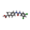  Mass: 383.226 Da / Num. of mol.: 2 / Source method: obtained synthetically / Formula: C17H16Cl2N2O4 Mass: 383.226 Da / Num. of mol.: 2 / Source method: obtained synthetically / Formula: C17H16Cl2N2O4#5: Water | ChemComp-HOH / | Water Mass: 18.015 Da / Num. of mol.: 220 / Source method: isolated from a natural source / Formula: H2O Mass: 18.015 Da / Num. of mol.: 220 / Source method: isolated from a natural source / Formula: H2O |
|---|
-Experimental details
-Experiment
| Experiment | Method: X-RAY DIFFRACTION / Number of used crystals: 1 |
|---|
- Sample preparation
| Crystal | Density Matthews: 2.16 Å3/Da / Density % sol: 42.98 % |
|---|---|
| Crystal grow | Temperature: 293 K / Method: batch / pH: 6.5 Details: 2.5M Ammonium sulfate, Ammonium phosphate, 0.1mM L35, pH 6.5, BATCH, temperature 293K |
-Data collection
| Diffraction | Mean temperature: 100 K |
|---|---|
| Diffraction source | Source: SYNCHROTRON / Site: SPring-8  / Beamline: BL40B2 / Beamline: BL40B2 |
| Detector | Type: ADSC QUANTUM 4 / Detector: CCD |
| Radiation | Protocol: SINGLE WAVELENGTH / Monochromatic (M) / Laue (L): M / Scattering type: x-ray |
| Radiation wavelength | Relative weight: 1 |
| Reflection | Resolution: 1.7→20 Å / Num. obs: 49000 / % possible obs: 85.1 % / Observed criterion σ(F): 0 / Observed criterion σ(I): 0 / Redundancy: 2.45 % / Rmerge(I) obs: 0.056 / Net I/σ(I): 13.1 |
| Reflection shell | Resolution: 1.7→1.76 Å / Rmerge(I) obs: 0.258 / % possible all: 63.8 |
- Processing
| Software |
| ||||||||||||||||||||||||||||||||||||||||||||||||||||||||||||||||||||||
|---|---|---|---|---|---|---|---|---|---|---|---|---|---|---|---|---|---|---|---|---|---|---|---|---|---|---|---|---|---|---|---|---|---|---|---|---|---|---|---|---|---|---|---|---|---|---|---|---|---|---|---|---|---|---|---|---|---|---|---|---|---|---|---|---|---|---|---|---|---|---|---|
| Refinement | Method to determine structure: MOLECULAR REPLACEMENT / Resolution: 1.7→19.88 Å / Cor.coef. Fo:Fc: 0.947 / Cor.coef. Fo:Fc free: 0.92 / SU B: 4.203 / SU ML: 0.124 / Cross valid method: THROUGHOUT / σ(F): 0 / ESU R: 0.143 / ESU R Free: 0.134 / Stereochemistry target values: MAXIMUM LIKELIHOOD
| ||||||||||||||||||||||||||||||||||||||||||||||||||||||||||||||||||||||
| Solvent computation | Ion probe radii: 0.8 Å / Shrinkage radii: 0.8 Å / VDW probe radii: 1.4 Å / Solvent model: BABINET MODEL WITH MASK | ||||||||||||||||||||||||||||||||||||||||||||||||||||||||||||||||||||||
| Displacement parameters | Biso mean: 17.804 Å2
| ||||||||||||||||||||||||||||||||||||||||||||||||||||||||||||||||||||||
| Refinement step | Cycle: LAST / Resolution: 1.7→19.88 Å
| ||||||||||||||||||||||||||||||||||||||||||||||||||||||||||||||||||||||
| Refine LS restraints |
| ||||||||||||||||||||||||||||||||||||||||||||||||||||||||||||||||||||||
| LS refinement shell | Resolution: 1.703→1.747 Å / Total num. of bins used: 20 /
|
