 Movie
Movie Controller

[English] 日本語
 Yorodumi
- PDB-2d5w: The crystal structure of oligopeptide binding protein from Thermu... -
+ Open data

- Basic information
| Entry | Database: PDB / ID: 2d5w | ||||||
|---|---|---|---|---|---|---|---|
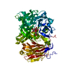
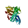
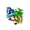
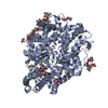
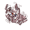
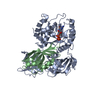
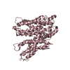
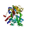
| Title | The crystal structure of oligopeptide binding protein from Thermus thermophilus HB8 complexed with pentapeptide | ||||||
 Components Components |
| ||||||
 Keywords Keywords | PEPTIDE BINDING PROTEIN / PROTEIN-PEPTIDE COMPLEX | ||||||
| Function / homology |  Function and homology information Function and homology informationATP-binding cassette (ABC) transporter complex / transmembrane transport Similarity search - Function | ||||||
| Biological species |    Thermus thermophilus (bacteria) Thermus thermophilus (bacteria) | ||||||
| Method | X-RAY DIFFRACTION / SYNCHROTRON / MAD / Resolution: 1.3 Å | ||||||
 Authors Authors | Morita, A. | ||||||
 Citation Citation | Journal: To be Published Title: The crystal structure of oligopeptide binding protein from Thermus thermophilus HB8 complexed with pentapeptide Authors: Morita, A. / Sakai, N. / Yao, M. / Watanabe, N. / Tanaka, I. | ||||||
| History |
|
- Structure visualization
| Structure viewer | Molecule: MolmilJmol/JSmol |
|---|
- Downloads & links
-Download
| PDBx/mmCIF format | 2d5w.cif.gz | 262.3 KB | Display | PDBx/mmCIF format |
|---|---|---|---|---|
| PDB format | pdb2d5w.ent.gz | 218.7 KB | Display | PDB format |
| PDBx/mmJSON format | 2d5w.json.gz | Tree view | PDBx/mmJSON format | |
| Others |  Other downloads Other downloads |
-Validation report
| Arichive directory | https://data.pdbj.org/pub/pdb/validation_reports/d5/2d5wftp://data.pdbj.org/pub/pdb/validation_reports/d5/2d5w | HTTPS FTP |
|---|
-Related structure data
| Similar structure data |
|---|


-Links
 PDBj
PDBj- Assembly
| Deposited unit | 
| ||||||||
|---|---|---|---|---|---|---|---|---|---|
| 1 | 
| ||||||||
| 2 | 
| ||||||||
| Unit cell |
|
-Components
| #1: Protein | Mass: 68704.859 Da / Num. of mol.: 2 / Fragment: residues 1-602 Source method: isolated from a genetically manipulated source Source: (gene. exp.) Thermus thermophilus (bacteria) / Strain: HB8 / Gene: TTHA1634 / Plasmid: pET-26b / Species (production host): Escherichia coli / Production host: Escherichia coli BL21(DE3) (bacteria) / Strain (production host): BL21(DE3) / References: UniProt: Q5SHU6#2: Protein/peptide | | Mass: 531.645 Da / Num. of mol.: 1 Source method: isolated from a genetically manipulated source Source: (gene. exp.) Thermus thermophilus (bacteria) / Strain: HB8Description: This peptide was co-purified and co-crystallized with the protein. Gene: TTHA1634 / Plasmid: pET-26b / Species (production host): Escherichia coli / Production host: Escherichia coli BL21(DE3) (bacteria) / Strain (production host): BL21(DE3)#3: Protein/peptide | | Mass: 535.634 Da / Num. of mol.: 1 Source method: isolated from a genetically manipulated source Source: (gene. exp.) Thermus thermophilus (bacteria) / Strain: HB8Description: This peptide was co-purified and co-crystallized with the protein. Gene: TTHA1634 / Plasmid: pET-26b / Species (production host): Escherichia coli / Production host: Escherichia coli BL21(DE3) (bacteria) / Strain (production host): BL21(DE3)#4: Water | ChemComp-HOH / | Water Mass: 18.015 Da / Num. of mol.: 1070 / Source method: isolated from a natural source / Formula: H2O Mass: 18.015 Da / Num. of mol.: 1070 / Source method: isolated from a natural source / Formula: H2O |
|---|
-Experimental details
-Experiment
| Experiment | Method: X-RAY DIFFRACTION / Number of used crystals: 2 |
|---|
- Sample preparation
| Crystal | Density Matthews: 2.28 Å3/Da / Density % sol: 45.98 % |
|---|---|
| Crystal grow | Temperature: 293 K / Method: vapor diffusion, hanging drop / pH: 7.5 Details: 20% PEG 3350, 0.15M lithium nitrate, pH 7.5, VAPOR DIFFUSION, HANGING DROP, temperature 293K |
-Data collection
| Diffraction |
| |||||||||||||||
|---|---|---|---|---|---|---|---|---|---|---|---|---|---|---|---|---|
| Diffraction source |
| |||||||||||||||
| Detector |
| |||||||||||||||
| Radiation |
| |||||||||||||||
| Radiation wavelength |
| |||||||||||||||
| Reflection | Resolution: 1.25→50 Å / Num. obs: 346050 / % possible obs: 100 % / Redundancy: 10.1 % / Biso Wilson estimate: 17.709 Å2 / Rsym value: 0.095 / Net I/σ(I): 31 | |||||||||||||||
| Reflection shell | Resolution: 1.25→1.29 Å / Redundancy: 6.7 % / Mean I/σ(I) obs: 6.3 / Rsym value: 0.412 / % possible all: 100 |

- Processing
| Software |
| |||||||||||||||||||||
|---|---|---|---|---|---|---|---|---|---|---|---|---|---|---|---|---|---|---|---|---|---|---|
| Refinement | Method to determine structure: MAD / Resolution: 1.3→15 Å / Cross valid method: THROUGHOUT / Stereochemistry target values: Engh & Huber
| |||||||||||||||||||||
| Displacement parameters | Biso mean: 11.527 Å2
| |||||||||||||||||||||
| Refine analyze |
| |||||||||||||||||||||
| Refinement step | Cycle: LAST / Resolution: 1.3→15 Å
| |||||||||||||||||||||
| Refine LS restraints |
| |||||||||||||||||||||
| LS refinement shell | Resolution: 1.3→1.35 Å
|
