 Movie
Movie Controller


+ Open data

- Basic information
| Entry | Database: PDB / ID: 2d58 | ||||||
|---|---|---|---|---|---|---|---|
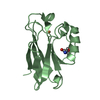
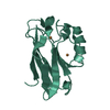
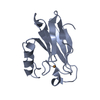
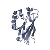
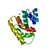
| Title | Human microglia-specific protein Iba1 | ||||||
 Components Components | Allograft inflammatory factor 1 | ||||||
 Keywords Keywords | METAL BINDING PROTEIN / EF-hand | ||||||
| Function / homology |  Function and homology information Function and homology informationnegative regulation of smooth muscle cell chemotaxis / positive regulation of smooth muscle cell chemotaxis / parallel actin filament bundle assembly / positive regulation of fibroblast growth factor production / actin crosslink formation / ruffle assembly / positive regulation of chemotaxis / positive regulation of mononuclear cell migration / positive regulation of monocyte chemotaxis / phagocytosis, engulfment ...negative regulation of smooth muscle cell chemotaxis / positive regulation of smooth muscle cell chemotaxis / parallel actin filament bundle assembly / positive regulation of fibroblast growth factor production / actin crosslink formation / ruffle assembly / positive regulation of chemotaxis / positive regulation of mononuclear cell migration / positive regulation of monocyte chemotaxis / phagocytosis, engulfment / Rac protein signal transduction / glial cell projection / phagocytic cup / actin filament bundle assembly / positive regulation of T cell migration / positive regulation of G1/S transition of mitotic cell cycle / positive regulation of T cell proliferation / positive regulation of chemokine production / ruffle / actin filament polymerization / actin filament / negative regulation of smooth muscle cell proliferation / positive regulation of smooth muscle cell proliferation / microglial cell activation / ruffle membrane / cellular response to type II interferon / positive regulation of interleukin-6 production / actin filament binding / lamellipodium / cellular response to oxidative stress / regulation of gene expression / positive regulation of cell migration / inflammatory response / calcium ion binding / positive regulation of cell population proliferation / perinuclear region of cytoplasm / nucleus / cytosol / cytoplasmSimilarity search - Function | ||||||
| Biological species |  Homo sapiens (human) Homo sapiens (human) | ||||||
| Method | X-RAY DIFFRACTION / SYNCHROTRON / MOLECULAR REPLACEMENT / Resolution: 1.9 Å | ||||||
 Authors Authors | Kamitori, S. | ||||||
 Citation Citation | Journal: J.Mol.Biol. / Year: 2006 Title: X-ray Structures of the Microglia/Macrophage-specific Protein Iba1 from Human and Mouse Demonstrate Novel Molecular Conformation Change Induced by Calcium binding Authors: Yamada, M. / Ohsawa, K. / Imai, Y. / Kohsaka, S. / Kamitori, S. | ||||||
| History |
|
- Structure visualization
| Structure viewer | Molecule: MolmilJmol/JSmol |
|---|
- Downloads & links
-Download
| PDBx/mmCIF format | 2d58.cif.gz | 36.3 KB | Display | PDBx/mmCIF format |
|---|---|---|---|---|
| PDB format | pdb2d58.ent.gz | 24.4 KB | Display | PDB format |
| PDBx/mmJSON format | 2d58.json.gz | Tree view | PDBx/mmJSON format | |
| Others |  Other downloads Other downloads |
-Validation report
| Arichive directory | https://data.pdbj.org/pub/pdb/validation_reports/d5/2d58ftp://data.pdbj.org/pub/pdb/validation_reports/d5/2d58 | HTTPS FTP |
|---|
-Related structure data


-Links
 PDBj
PDBj- Assembly
| Deposited unit | 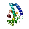
| ||||||||
|---|---|---|---|---|---|---|---|---|---|
| 1 |
| ||||||||
| Unit cell |
|
-Components
| #1: Protein | / AIF-1 / Ionized calcium-binding adapter molecule 1 / G1 protein Mass: 12338.285 Da / Num. of mol.: 1 / Fragment: residues 17-123 Source method: isolated from a genetically manipulated source Source: (gene. exp.) Homo sapiens (human) / Production host:  Escherichia coli (E. coli) / References: UniProt: P55008 Escherichia coli (E. coli) / References: UniProt: P55008 |
|---|---|
| #2: Chemical | ChemComp-NI / Nickel  Mass: 58.693 Da / Num. of mol.: 1 / Source method: obtained synthetically / Formula: Ni Mass: 58.693 Da / Num. of mol.: 1 / Source method: obtained synthetically / Formula: Ni |
| #3: Water | ChemComp-HOH / Water Mass: 18.015 Da / Num. of mol.: 107 / Source method: isolated from a natural source / Formula: H2O Mass: 18.015 Da / Num. of mol.: 107 / Source method: isolated from a natural source / Formula: H2O |
-Experimental details
-Experiment
| Experiment | Method: X-RAY DIFFRACTION / Number of used crystals: 1 |
|---|
- Sample preparation
| Crystal | Density Matthews: 1.94 Å3/Da / Density % sol: 36.66 % |
|---|---|
| Crystal grow | Temperature: 300 K / Method: vapor diffusion / pH: 7.3 Details: 20% (w/v) polyethylene glycol (PEG) 4000, 10% (w/v) 2-propanol, 0.01M NiCl2 in 100mM of Tris-HCl (pH 7.3), VAPOR DIFFUSION, temperature 300K |
-Data collection
| Diffraction | Mean temperature: 100 K |
|---|---|
| Diffraction source | Source: SYNCHROTRON / Site: Photon Factory  / Beamline: BL-6A / Wavelength: 1 Å / Beamline: BL-6A / Wavelength: 1 Å |
| Detector | Type: ADSC QUANTUM 4 / Detector: CCD / Date: Jun 28, 2004 |
| Radiation | Monochromator: graphite / Protocol: SINGLE WAVELENGTH / Monochromatic (M) / Laue (L): M / Scattering type: x-ray |
| Radiation wavelength | Wavelength: 1 Å / Relative weight: 1 |
| Reflection | Resolution: 1.9→31.24 Å / Num. all: 7462 / Num. obs: 7462 / % possible obs: 97.4 % / Observed criterion σ(F): 0 / Observed criterion σ(I): 0 / Biso Wilson estimate: 12.6 Å2 / Rmerge(I) obs: 0.76 |
| Reflection shell | Resolution: 1.9→2.02 Å / % possible all: 90.1 |
- Processing
| Software |
| ||||||||||||||||||||||||||||||||||||||||||||||||||||||||||||||||||||||||||||||||
|---|---|---|---|---|---|---|---|---|---|---|---|---|---|---|---|---|---|---|---|---|---|---|---|---|---|---|---|---|---|---|---|---|---|---|---|---|---|---|---|---|---|---|---|---|---|---|---|---|---|---|---|---|---|---|---|---|---|---|---|---|---|---|---|---|---|---|---|---|---|---|---|---|---|---|---|---|---|---|---|---|---|
| Refinement | Method to determine structure: MOLECULAR REPLACEMENT / Resolution: 1.9→31.24 Å / Rfactor Rfree error: 0.009 / Data cutoff high absF: 116478.66 / Data cutoff low absF: 0 / Isotropic thermal model: RESTRAINED / Cross valid method: THROUGHOUT / σ(F): 0 / Stereochemistry target values: Engh & Huber
| ||||||||||||||||||||||||||||||||||||||||||||||||||||||||||||||||||||||||||||||||
| Solvent computation | Solvent model: FLAT MODEL / Bsol: 52.6003 Å2 / ksol: 0.350076 e/Å3 | ||||||||||||||||||||||||||||||||||||||||||||||||||||||||||||||||||||||||||||||||
| Displacement parameters | Biso mean: 20.7 Å2
| ||||||||||||||||||||||||||||||||||||||||||||||||||||||||||||||||||||||||||||||||
| Refine analyze |
| ||||||||||||||||||||||||||||||||||||||||||||||||||||||||||||||||||||||||||||||||
| Refinement step | Cycle: LAST / Resolution: 1.9→31.24 Å
| ||||||||||||||||||||||||||||||||||||||||||||||||||||||||||||||||||||||||||||||||
| Refine LS restraints |
| ||||||||||||||||||||||||||||||||||||||||||||||||||||||||||||||||||||||||||||||||
| LS refinement shell | Resolution: 1.9→2.02 Å / Total num. of bins used: 6
| ||||||||||||||||||||||||||||||||||||||||||||||||||||||||||||||||||||||||||||||||
| Xplor file |
|
