 Movie
Movie Controller

[English] 日本語
 Yorodumi
- PDB-2d4d: The Crystal Structure of human beta2-microglobulin, L39W W60F W95... -
+ Open data

- Basic information
| Entry | Database: PDB / ID: 2d4d | ||||||
|---|---|---|---|---|---|---|---|
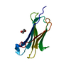
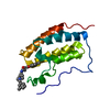
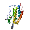
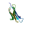
| Title | The Crystal Structure of human beta2-microglobulin, L39W W60F W95F Mutant | ||||||
 Components Components | Beta-2-microglobulin Beta-2 microglobulin Beta-2 microglobulin | ||||||
 Keywords Keywords | IMMUNE SYSTEM / Immunoglobulin constant domain | ||||||
| Function / homology |  Function and homology information Function and homology informationpositive regulation of ferrous iron binding / positive regulation of transferrin receptor binding / positive regulation of receptor binding / early endosome lumen / Nef mediated downregulation of MHC class I complex cell surface expression / DAP12 interactions / negative regulation of receptor binding / Endosomal/Vacuolar pathway / Antigen Presentation: Folding, assembly and peptide loading of class I MHC / cellular response to iron(III) ion ...positive regulation of ferrous iron binding / positive regulation of transferrin receptor binding / positive regulation of receptor binding / early endosome lumen / Nef mediated downregulation of MHC class I complex cell surface expression / DAP12 interactions / negative regulation of receptor binding / Endosomal/Vacuolar pathway / Antigen Presentation: Folding, assembly and peptide loading of class I MHC / cellular response to iron(III) ion / antigen processing and presentation of exogenous protein antigen via MHC class Ib, TAP-dependent / negative regulation of forebrain neuron differentiation / ER to Golgi transport vesicle membrane / regulation of erythrocyte differentiation / peptide antigen assembly with MHC class I protein complex / response to molecule of bacterial origin / regulation of iron ion transport / MHC class I peptide loading complex / HFE-transferrin receptor complex / T cell mediated cytotoxicity / cellular response to iron ion / antigen processing and presentation of endogenous peptide antigen via MHC class I / positive regulation of T cell cytokine production / MHC class I protein complex / multicellular organismal-level iron ion homeostasis / negative regulation of neurogenesis / peptide antigen assembly with MHC class II protein complex / positive regulation of receptor-mediated endocytosis / positive regulation of T cell mediated cytotoxicity / MHC class II protein complex / cellular response to nicotine / recycling endosome membrane / specific granule lumen / phagocytic vesicle membrane / peptide antigen binding / positive regulation of cellular senescence / antigen processing and presentation of exogenous peptide antigen via MHC class II / negative regulation of epithelial cell proliferation / Immunoregulatory interactions between a Lymphoid and a non-Lymphoid cell / Interferon gamma signaling / positive regulation of immune response / Modulation by Mtb of host immune system / sensory perception of smell / positive regulation of T cell activation / positive regulation of protein binding / tertiary granule lumen / DAP12 signaling / negative regulation of neuron projection development / MHC class II protein complex binding / late endosome membrane / T cell differentiation in thymus / ER-Phagosome pathway / iron ion transport / early endosome membrane / protein refolding / protein homotetramerization / intracellular iron ion homeostasis / amyloid fibril formation / learning or memory / Amyloid fiber formation / lysosomal membrane / external side of plasma membrane / endoplasmic reticulum lumen / Golgi membrane / focal adhesion / Neutrophil degranulation / SARS-CoV-2 activates/modulates innate and adaptive immune responses / structural molecule activity / Golgi apparatus / endoplasmic reticulum / protein homodimerization activity / extracellular space / extracellular exosome / extracellular region / membrane / identical protein binding / plasma membrane / cytosolSimilarity search - Function | ||||||
| Biological species |  Homo sapiens (human) Homo sapiens (human) | ||||||
| Method | X-RAY DIFFRACTION / MOLECULAR REPLACEMENT / Resolution: 2.1 Å | ||||||
 Authors Authors | Iwata, K. / Matsuura, T. / Nakagawa, A. / Goto, Y. | ||||||
 Citation Citation | Journal: J.Biol.Chem. / Year: 2006 Title: Conformation of Amyloid Fibrils of beta2-Microglobulin Probed by Tryptophan Mutagenesis Authors: Kihara, M. / Chatani, E. / Iwata, K. / Yamamoto, K. / Matsuura, T. / Nakagawa, A. / Naiki, H. / Goto, Y. | ||||||
| History |
|
- Structure visualization
| Structure viewer | Molecule: MolmilJmol/JSmol |
|---|
- Downloads & links
-Download
| PDBx/mmCIF format | 2d4d.cif.gz | 34.9 KB | Display | PDBx/mmCIF format |
|---|---|---|---|---|
| PDB format | pdb2d4d.ent.gz | 22.2 KB | Display | PDB format |
| PDBx/mmJSON format | 2d4d.json.gz | Tree view | PDBx/mmJSON format | |
| Others |  Other downloads Other downloads |
-Validation report
| Arichive directory | https://data.pdbj.org/pub/pdb/validation_reports/d4/2d4dftp://data.pdbj.org/pub/pdb/validation_reports/d4/2d4d | HTTPS FTP |
|---|
-Related structure data
| Related structure data | 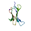 2d4fC 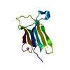 1ldsS C: citing same article ( S: Starting model for refinement |
|---|---|
| Similar structure data |


-Links
 PDBj
PDBj

- Assembly
| Deposited unit | 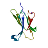
| ||||||||
|---|---|---|---|---|---|---|---|---|---|
| 1 |
| ||||||||
| Unit cell |
|
-Components
| #1: Protein | Beta-2 microglobulin Mass: 11874.338 Da / Num. of mol.: 1 / Mutation: L39W, W60F, W95F Source method: isolated from a genetically manipulated source Source: (gene. exp.) Homo sapiens (human) / Plasmid: pHIKARU1a / Production host:  Escherichia coli (E. coli) / Strain (production host): BL21 (DE3) pLysS / References: UniProt: P61769 Escherichia coli (E. coli) / Strain (production host): BL21 (DE3) pLysS / References: UniProt: P61769 |
|---|---|
| #2: Chemical | ChemComp-NA /   Mass: 22.990 Da / Num. of mol.: 1 / Source method: obtained synthetically / Formula: Na Mass: 22.990 Da / Num. of mol.: 1 / Source method: obtained synthetically / Formula: Na |
| #3: Water | ChemComp-HOH / Water Mass: 18.015 Da / Num. of mol.: 48 / Source method: isolated from a natural source / Formula: H2O Mass: 18.015 Da / Num. of mol.: 48 / Source method: isolated from a natural source / Formula: H2O |
-Experimental details
-Experiment
| Experiment | Method: X-RAY DIFFRACTION / Number of used crystals: 1 |
|---|
- Sample preparation
| Crystal | Density Matthews: 2.24 Å3/Da / Density % sol: 45 % |
|---|---|
| Crystal grow | Temperature: 288 K / Method: vapor diffusion, hanging drop / pH: 5.6 Details: 15% PEG4000, 25% glycerol, 0.2M ammonium acetate, 0.1M sodium acetate, pH 5.6, VAPOR DIFFUSION, HANGING DROP, temperature 288K |
-Data collection
| Diffraction | Mean temperature: 100 K |
|---|---|
| Diffraction source | Source: ROTATING ANODE / Type: RIGAKU FR-E / Wavelength: 1.5418 Å |
| Detector | Type: RIGAKU RAXIS VII / Detector: IMAGE PLATE / Date: Jun 24, 2005 |
| Radiation | Monochromator: conforcal mirror / Protocol: SINGLE WAVELENGTH / Monochromatic (M) / Laue (L): M / Scattering type: x-ray |
| Radiation wavelength | Wavelength: 1.5418 Å / Relative weight: 1 |
| Reflection | Resolution: 2.1→47.673 Å / Num. all: 6345 / Num. obs: 6343 / % possible obs: 100 % / Observed criterion σ(F): 3 / Redundancy: 3.5 % / Biso Wilson estimate: 26.44 Å2 / Rmerge(I) obs: 0.076 / Net I/σ(I): 8.2 |
| Reflection shell | Resolution: 2.1→2.21 Å / Redundancy: 927 % / Mean I/σ(I) obs: 3.5 / Rsym value: 0.315 / % possible all: 100 |
- Processing
| Software |
| |||||||||||||||||||||||||||||||||||||||||||||||||||||||||||||||||||||||||||||||||||||||||||||||
|---|---|---|---|---|---|---|---|---|---|---|---|---|---|---|---|---|---|---|---|---|---|---|---|---|---|---|---|---|---|---|---|---|---|---|---|---|---|---|---|---|---|---|---|---|---|---|---|---|---|---|---|---|---|---|---|---|---|---|---|---|---|---|---|---|---|---|---|---|---|---|---|---|---|---|---|---|---|---|---|---|---|---|---|---|---|---|---|---|---|---|---|---|---|---|---|---|
| Refinement | Method to determine structure: MOLECULAR REPLACEMENT Starting model: PDB entry 1LDS Resolution: 2.1→47.67 Å / Cor.coef. Fo:Fc: 0.935 / Cor.coef. Fo:Fc free: 0.909 / SU B: 5.948 / SU ML: 0.157 / Cross valid method: THROUGHOUT / σ(F): 0 / ESU R: 0.262 / ESU R Free: 0.213 / Stereochemistry target values: MAXIMUM LIKELIHOOD
| |||||||||||||||||||||||||||||||||||||||||||||||||||||||||||||||||||||||||||||||||||||||||||||||
| Solvent computation | Ion probe radii: 0.8 Å / Shrinkage radii: 0.8 Å / VDW probe radii: 1.2 Å / Solvent model: MASK | |||||||||||||||||||||||||||||||||||||||||||||||||||||||||||||||||||||||||||||||||||||||||||||||
| Displacement parameters | Biso mean: 25.258 Å2
| |||||||||||||||||||||||||||||||||||||||||||||||||||||||||||||||||||||||||||||||||||||||||||||||
| Refinement step | Cycle: LAST / Resolution: 2.1→47.67 Å
| |||||||||||||||||||||||||||||||||||||||||||||||||||||||||||||||||||||||||||||||||||||||||||||||
| Refine LS restraints |
| |||||||||||||||||||||||||||||||||||||||||||||||||||||||||||||||||||||||||||||||||||||||||||||||
| LS refinement shell | Resolution: 2.1→2.155 Å / Total num. of bins used: 20
|
