 Movie
Movie Controller


+ Open data

- Basic information
| Entry | Database: PDB / ID: 2d1i | ||||||
|---|---|---|---|---|---|---|---|
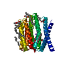
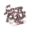
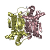
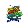
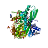
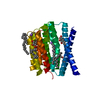
| Title | Structure of human Atg4b | ||||||
 Components Components | Cysteine protease APG4B | ||||||
 Keywords Keywords |  HYDROLASE / CYSTEINE PROTEASE / AUTOPHAGY / ATG / APG HYDROLASE / CYSTEINE PROTEASE / AUTOPHAGY / ATG / APG | ||||||
| Function / homology |  Function and homology information Function and homology informationprotein-phosphatidylethanolamide deconjugating activity / otolith mineralization completed early in development / protein delipidation / microautophagy / protein localization to phagophore assembly site / Macroautophagy / autophagosome membrane / mitophagy / autophagosome assembly / cysteine-type peptidase activity ...protein-phosphatidylethanolamide deconjugating activity / otolith mineralization completed early in development / protein delipidation / microautophagy / protein localization to phagophore assembly site / Macroautophagy / autophagosome membrane / mitophagy / autophagosome assembly / cysteine-type peptidase activity / macroautophagy / autophagy / protein transport / cytoplasmic vesicle / scaffold protein binding / endopeptidase activity / Hydrolases; Acting on peptide bonds (peptidases); Cysteine endopeptidases / cysteine-type endopeptidase activity / endoplasmic reticulum / mitochondrion / proteolysis / cytosolSimilarity search - Function | ||||||
| Biological species |  Homo sapiens (human) Homo sapiens (human) | ||||||
| Method | X-RAY DIFFRACTION / SYNCHROTRON / MIR / Resolution: 2 Å | ||||||
 Authors Authors | Kumanomidou, T. / Mizushima, T. / Komatsu, M. / Suzuki, A. / Tanida, I. / Sou, Y.S. / Ueno, T. / Kominami, E. / Tanaka, K. / Yamane, T. | ||||||
 Citation Citation | Journal: J.Mol.Biol. / Year: 2006 Title: The Crystal Structure of Human Atg4b, a Processing and De-conjugating Enzyme for Autophagosome-forming Modifiers Authors: Kumanomidou, T. / Mizushima, T. / Komatsu, M. / Suzuki, A. / Tanida, I. / Sou, Y.S. / Ueno, T. / Kominami, E. / Tanaka, K. / Yamane, T. | ||||||
| History |
|
- Structure visualization
| Structure viewer | Molecule: MolmilJmol/JSmol |
|---|
- Downloads & links
-Download
| PDBx/mmCIF format | 2d1i.cif.gz | 152.5 KB | Display | PDBx/mmCIF format |
|---|---|---|---|---|
| PDB format | pdb2d1i.ent.gz | 119.8 KB | Display | PDB format |
| PDBx/mmJSON format | 2d1i.json.gz | Tree view | PDBx/mmJSON format | |
| Others |  Other downloads Other downloads |
-Validation report
| Arichive directory | https://data.pdbj.org/pub/pdb/validation_reports/d1/2d1iftp://data.pdbj.org/pub/pdb/validation_reports/d1/2d1i | HTTPS FTP |
|---|
-Related structure data
| Similar structure data |
|---|


-Links
 PDBj
PDBj
- Assembly
| Deposited unit | 
| ||||||||
|---|---|---|---|---|---|---|---|---|---|
| 1 | 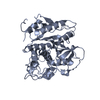
| ||||||||
| 2 | 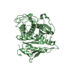
| ||||||||
| Unit cell |
|
-Components
| #1: Protein | Mass: 44764.625 Da / Num. of mol.: 2 Source method: isolated from a genetically manipulated source Source: (gene. exp.) Homo sapiens (human) / Plasmid: pGEX6P / Species (production host): Escherichia coli / Production host:  Escherichia coli BL21(DE3) (bacteria) / Strain (production host): BL21(DE3) Escherichia coli BL21(DE3) (bacteria) / Strain (production host): BL21(DE3)References: UniProt: Q9Y4P1, Hydrolases; Acting on peptide bonds (peptidases); Cysteine endopeptidases#2: Water | ChemComp-HOH / | Water Mass: 18.015 Da / Num. of mol.: 338 / Source method: isolated from a natural source / Formula: H2O Mass: 18.015 Da / Num. of mol.: 338 / Source method: isolated from a natural source / Formula: H2O |
|---|
-Experimental details
-Experiment
| Experiment | Method: X-RAY DIFFRACTION / Number of used crystals: 7 |
|---|
- Sample preparation
| Crystal | Density Matthews: 2.1 Å3/Da / Density % sol: 40.4 % |
|---|---|
| Crystal grow | Temperature: 288 K / Method: vapor diffusion, hanging drop / pH: 6.5 Details: 0.65M tri-Sodium citrate dihydrate, pH 6.5, VAPOR DIFFUSION, HANGING DROP, temperature 288K |
-Data collection
| Diffraction | Mean temperature: 100 K |
|---|---|
| Diffraction source | Source: SYNCHROTRON / Site: SPring-8  / Beamline: BL44XU / Wavelength: 0.9 Å / Beamline: BL44XU / Wavelength: 0.9 Å |
| Detector | Type: Bruker DIP-6040 / Detector: CCD / Date: Oct 9, 2004 |
| Radiation | Protocol: SINGLE WAVELENGTH / Monochromatic (M) / Laue (L): M / Scattering type: x-ray |
| Radiation wavelength | Wavelength: 0.9 Å / Relative weight: 1 |
| Reflection | Resolution: 2→44.59 Å / Num. obs: 48166 / % possible obs: 98.7 % / Observed criterion σ(I): -3 / Redundancy: 3.7 % / Biso Wilson estimate: 33.018 Å2 |
| Reflection shell | Resolution: 2→2.07 Å / % possible all: 99.1 |
- Processing
| Software |
| ||||||||||||||||||||||||||||||||||||||||||||||||||||||||||||||||||||||||||||||||||||||||||
|---|---|---|---|---|---|---|---|---|---|---|---|---|---|---|---|---|---|---|---|---|---|---|---|---|---|---|---|---|---|---|---|---|---|---|---|---|---|---|---|---|---|---|---|---|---|---|---|---|---|---|---|---|---|---|---|---|---|---|---|---|---|---|---|---|---|---|---|---|---|---|---|---|---|---|---|---|---|---|---|---|---|---|---|---|---|---|---|---|---|---|---|
| Refinement | Method to determine structure: MIR / Resolution: 2→44.59 Å / Cor.coef. Fo:Fc: 0.945 / Cor.coef. Fo:Fc free: 0.908 / SU B: 4.916 / SU ML: 0.141 / Cross valid method: THROUGHOUT / ESU R: 0.226 / ESU R Free: 0.205 / Stereochemistry target values: MAXIMUM LIKELIHOOD / Details: HYDROGENS HAVE BEEN ADDED IN THE RIDING POSITIONS
| ||||||||||||||||||||||||||||||||||||||||||||||||||||||||||||||||||||||||||||||||||||||||||
| Solvent computation | Ion probe radii: 0.8 Å / Shrinkage radii: 0.8 Å / VDW probe radii: 1.2 Å / Solvent model: MASK | ||||||||||||||||||||||||||||||||||||||||||||||||||||||||||||||||||||||||||||||||||||||||||
| Displacement parameters | Biso mean: 40.978 Å2
| ||||||||||||||||||||||||||||||||||||||||||||||||||||||||||||||||||||||||||||||||||||||||||
| Refinement step | Cycle: LAST / Resolution: 2→44.59 Å
| ||||||||||||||||||||||||||||||||||||||||||||||||||||||||||||||||||||||||||||||||||||||||||
| Refine LS restraints |
| ||||||||||||||||||||||||||||||||||||||||||||||||||||||||||||||||||||||||||||||||||||||||||
| LS refinement shell | Resolution: 2→2.052 Å / Total num. of bins used: 20
|
