 Movie
Movie Controller


+ Open data

- Basic information
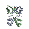
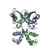
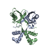
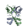
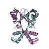
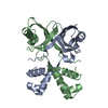
| Entry | Database: PDB / ID: 2cyy | ||||||
|---|---|---|---|---|---|---|---|
| Title | Crystal structure of PH1519 from Pyrococcus horikosii OT3 | ||||||
 Components Components | Putative HTH-type transcriptional regulator PH1519 | ||||||
 Keywords Keywords |  DNA BINDING PROTEIN / structural genomics / Pyrococcus horikosii OT3 / NPPSFA / National Project on Protein Structural and Functional Analyses / RIKEN Structural Genomics/Proteomics Initiative / RSGI DNA BINDING PROTEIN / structural genomics / Pyrococcus horikosii OT3 / NPPSFA / National Project on Protein Structural and Functional Analyses / RIKEN Structural Genomics/Proteomics Initiative / RSGI | ||||||
| Function / homology |  Function and homology information Function and homology information | ||||||
| Biological species |   Pyrococcus horikoshii (archaea) Pyrococcus horikoshii (archaea) | ||||||
| Method | X-RAY DIFFRACTION / SYNCHROTRON / MAD / Resolution: 1.8 Å | ||||||
 Authors Authors | Okazaki, N. / Nakano, N. / Shinkai, A. / Yokoyama, S. / RIKEN Structural Genomics/Proteomics Initiative (RSGI) | ||||||
 Citation Citation | Journal: To be Published Title: Crystal structure of PH1519 from Pyrococcus horikosii OT3 Authors: Okazaki, N. / Nakano, N. / Shinkai, A. / Yokoyama, S. | ||||||
| History |
|
- Structure visualization
| Structure viewer | Molecule: MolmilJmol/JSmol |
|---|
- Downloads & links
-Download
| PDBx/mmCIF format | 2cyy.cif.gz | 45.8 KB | Display | PDBx/mmCIF format |
|---|---|---|---|---|
| PDB format | pdb2cyy.ent.gz | 35.6 KB | Display | PDB format |
| PDBx/mmJSON format | 2cyy.json.gz | Tree view | PDBx/mmJSON format | |
| Others |  Other downloads Other downloads |
-Validation report
| Arichive directory | https://data.pdbj.org/pub/pdb/validation_reports/cy/2cyyftp://data.pdbj.org/pub/pdb/validation_reports/cy/2cyy | HTTPS FTP |
|---|
-Related structure data
| Similar structure data | |
|---|---|
| Other databases |


-Links
 PDBj
PDBj- Assembly
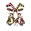
| Deposited unit | 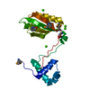
| ||||||||||||||||||||||||||||||||||||
|---|---|---|---|---|---|---|---|---|---|---|---|---|---|---|---|---|---|---|---|---|---|---|---|---|---|---|---|---|---|---|---|---|---|---|---|---|---|
| 1 | 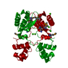
| ||||||||||||||||||||||||||||||||||||
| 2 |
| ||||||||||||||||||||||||||||||||||||
| Unit cell |
| ||||||||||||||||||||||||||||||||||||
| Components on special symmetry positions |
| ||||||||||||||||||||||||||||||||||||
| Details | The second part of the biological assembly is generated by the two fold axis: -x+y, y,-z+1/2. |
-Components
| #1: Protein | Mass: 17167.434 Da / Num. of mol.: 1 Source method: isolated from a genetically manipulated source Source: (gene. exp.) Pyrococcus horikoshii (archaea) / Plasmid: pET11a / Production host:  Escherichia coli (E. coli) / Strain (production host): BL21-CodonPlus(DE3)-RIL-X / References: UniProt: O59188 Escherichia coli (E. coli) / Strain (production host): BL21-CodonPlus(DE3)-RIL-X / References: UniProt: O59188 | ||||
|---|---|---|---|---|---|
| #2: Chemical | ChemComp-CA /   Mass: 40.078 Da / Num. of mol.: 4 / Source method: obtained synthetically / Formula: Ca Mass: 40.078 Da / Num. of mol.: 4 / Source method: obtained synthetically / Formula: Ca#3: Chemical | ChemComp-GLN / | Glutamine  Type: L-peptide linking / Mass: 146.144 Da / Num. of mol.: 1 / Source method: obtained synthetically / Formula: C5H10N2O3 Type: L-peptide linking / Mass: 146.144 Da / Num. of mol.: 1 / Source method: obtained synthetically / Formula: C5H10N2O3#4: Water | ChemComp-HOH / | Water Mass: 18.015 Da / Num. of mol.: 214 / Source method: isolated from a natural source / Formula: H2O Mass: 18.015 Da / Num. of mol.: 214 / Source method: isolated from a natural source / Formula: H2O |
-Experimental details
-Experiment
| Experiment | Method: X-RAY DIFFRACTION / Number of used crystals: 1 |
|---|
- Sample preparation
| Crystal | Density Matthews: 3.42 Å3/Da / Density % sol: 64.02 % |
|---|---|
| Crystal grow | Temperature: 291 K / Method: microbatch / pH: 8.5 Details: 25.5% PEG4000, 0.085M Tris-HCl, 0.17M Li2(SO4), 15% Glycerol, pH 8.5, Microbatch, temperature 291K |
-Data collection
| Diffraction | Mean temperature: 100 K | ||||||||||||
|---|---|---|---|---|---|---|---|---|---|---|---|---|---|
| Diffraction source | Source: SYNCHROTRON / Site: SPring-8  / Beamline: BL26B2 / Wavelength: 0.97901, 0.97940, 0.96000 / Beamline: BL26B2 / Wavelength: 0.97901, 0.97940, 0.96000 | ||||||||||||
| Detector | Type: RIGAKU JUPITER 210 / Detector: CCD / Date: Oct 26, 2004 / Details: mirrors | ||||||||||||
| Radiation | Monochromator: Bending Magnet / Protocol: MAD / Monochromatic (M) / Laue (L): M / Scattering type: x-ray | ||||||||||||
| Radiation wavelength |
| ||||||||||||
| Reflection | Resolution: 1.8→30 Å / Num. all: 22776 / Num. obs: 22776 / % possible obs: 100 % / Observed criterion σ(F): 0 / Observed criterion σ(I): 0 / Redundancy: 23.1 % / Biso Wilson estimate: 18.612 Å2 / Rmerge(I) obs: 0.076 / Rsym value: 0.062 / Net I/σ(I): 17.1 | ||||||||||||
| Reflection shell | Resolution: 1.8→1.86 Å / Redundancy: 21.9 % / Rmerge(I) obs: 0.354 / Mean I/σ(I) obs: 8.84 / Num. unique all: 2239 / Rsym value: 0.341 / % possible all: 100 |
- Processing
| Software |
| |||||||||||||||||||||||||
|---|---|---|---|---|---|---|---|---|---|---|---|---|---|---|---|---|---|---|---|---|---|---|---|---|---|---|
| Refinement | Method to determine structure: MAD / Resolution: 1.8→29.58 Å / Isotropic thermal model: RESTRAINED / Cross valid method: THROUGHPUT / σ(F): 0 / Stereochemistry target values: Engh & Huber
| |||||||||||||||||||||||||
| Displacement parameters | Biso mean: 26.2 Å2
| |||||||||||||||||||||||||
| Refine analyze |
| |||||||||||||||||||||||||
| Refinement step | Cycle: LAST / Resolution: 1.8→29.58 Å
| |||||||||||||||||||||||||
| Refine LS restraints |
| |||||||||||||||||||||||||
| LS refinement shell | Resolution: 1.8→1.88 Å / Rfactor Rfree error: 0.02
|
