 Movie
Movie Controller

[English] 日本語
 Yorodumi
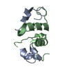
- PDB-2cwr: Crystal structure of chitin biding domain of chitinase from Pyroc... -
+ Open data

- Basic information
| Entry | Database: PDB / ID: 2cwr | ||||||
|---|---|---|---|---|---|---|---|
| Title | Crystal structure of chitin biding domain of chitinase from Pyrococcus furiosus | ||||||
 Components Components | chitinase | ||||||
 Keywords Keywords | HYDROLASE / chitinase / chitin-binding domain / Pyrococcus furiosus / endoglucanase / chitin / hyperthermophilic | ||||||
| Function / homology |  Function and homology informationpolysaccharide binding / chitin binding / hydrolase activity, hydrolyzing O-glycosyl compounds / carbohydrate metabolic process Function and homology informationpolysaccharide binding / chitin binding / hydrolase activity, hydrolyzing O-glycosyl compounds / carbohydrate metabolic processSimilarity search - Function | ||||||
| Biological species |   Pyrococcus furiosus (archaea) Pyrococcus furiosus (archaea) | ||||||
| Method | X-RAY DIFFRACTION / SYNCHROTRON / MOLECULAR REPLACEMENT / Resolution: 1.7 Å | ||||||
 Authors Authors | Uegaki, K. / Nakamura, T. / Ishikawa, K. / Matsumura, H. | ||||||
 Citation Citation | Journal: J.Mol.Biol. / Year: 2008 Title: Tertiary structure and carbohydrate recognition by the chitin-binding domain of a hyperthermophilic chitinase from Pyrococcus furiosus. Authors: Nakamura, T. / Mine, S. / Hagihara, Y. / Ishikawa, K. / Ikegami, T. / Uegaki, K. | ||||||
| History |
|
- Structure visualization
| Structure viewer | Molecule: MolmilJmol/JSmol |
|---|
- Downloads & links
-Download
| PDBx/mmCIF format | 2cwr.cif.gz | 32.6 KB | Display | PDBx/mmCIF format |
|---|---|---|---|---|
| PDB format | pdb2cwr.ent.gz | 22.2 KB | Display | PDB format |
| PDBx/mmJSON format | 2cwr.json.gz | Tree view | PDBx/mmJSON format | |
| Others |  Other downloads Other downloads |
-Validation report
| Arichive directory | https://data.pdbj.org/pub/pdb/validation_reports/cw/2cwrftp://data.pdbj.org/pub/pdb/validation_reports/cw/2cwr | HTTPS FTP |
|---|
-Related structure data


-Links
 PDBj
PDBj
- Assembly
| Deposited unit | 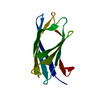
| ||||||||
|---|---|---|---|---|---|---|---|---|---|
| 1 |
| ||||||||
| Unit cell |
|
-Components
| #1: Protein | Mass: 10964.104 Da / Num. of mol.: 1 / Fragment: chitin-binding domain Source method: isolated from a genetically manipulated source Source: (gene. exp.) Pyrococcus furiosus (archaea) / Gene: PF1233 / Production host:  Escherichia coli (E. coli) / References: UniProt: Q8U1H5, chitinase Escherichia coli (E. coli) / References: UniProt: Q8U1H5, chitinase |
|---|---|
| #2: Water | ChemComp-HOH / Water Mass: 18.015 Da / Num. of mol.: 141 / Source method: isolated from a natural source / Formula: H2O Mass: 18.015 Da / Num. of mol.: 141 / Source method: isolated from a natural source / Formula: H2O |
-Experimental details
-Experiment
| Experiment | Method: X-RAY DIFFRACTION / Number of used crystals: 1 |
|---|
- Sample preparation
| Crystal | Density Matthews: 2.3 Å3/Da / Density % sol: 47 % |
|---|---|
| Crystal grow | Temperature: 298 K / Method: vapor diffusion, hanging drop / pH: 6.5 Details: PEG 8000, magnesium acetate, sodium cacodylate, pH 6.5, VAPOR DIFFUSION, HANGING DROP, temperature 298K |
-Data collection
| Diffraction | Mean temperature: 100 K |
|---|---|
| Diffraction source | Source: SYNCHROTRON / Site: SPring-8  / Beamline: BL38B1 / Wavelength: 1 Å / Beamline: BL38B1 / Wavelength: 1 Å |
| Detector | Type: RIGAKU JUPITER / Detector: CCD / Date: Oct 14, 2004 |
| Radiation | Protocol: SINGLE WAVELENGTH / Monochromatic (M) / Laue (L): M / Scattering type: x-ray |
| Radiation wavelength | Wavelength: 1 Å / Relative weight: 1 |
| Reflection | Resolution: 1.7→50 Å / Num. obs: 11986 / % possible obs: 96.8 % / Observed criterion σ(I): 14.2 / Redundancy: 12.7 % / Biso Wilson estimate: 14.3 Å2 / Rmerge(I) obs: 0.076 |
| Reflection shell | Resolution: 1.7→1.76 Å / Rmerge(I) obs: 0.233 / Mean I/σ(I) obs: 3 / % possible all: 80.5 |
- Processing
| Software |
| ||||||||||||||||||||||||||||||||||||
|---|---|---|---|---|---|---|---|---|---|---|---|---|---|---|---|---|---|---|---|---|---|---|---|---|---|---|---|---|---|---|---|---|---|---|---|---|---|
| Refinement | Method to determine structure: MOLECULAR REPLACEMENT / Resolution: 1.7→32.06 Å / Rfactor Rfree error: 0.007 / Data cutoff high absF: 1258850.71 / Data cutoff low absF: 0 / Isotropic thermal model: RESTRAINED / Cross valid method: THROUGHOUT / σ(F): 0 / Stereochemistry target values: Engh & Huber
| ||||||||||||||||||||||||||||||||||||
| Solvent computation | Solvent model: FLAT MODEL / Bsol: 39.9514 Å2 / ksol: 0.400825 e/Å3 | ||||||||||||||||||||||||||||||||||||
| Displacement parameters | Biso mean: 21.5 Å2
| ||||||||||||||||||||||||||||||||||||
| Refine analyze |
| ||||||||||||||||||||||||||||||||||||
| Refinement step | Cycle: LAST / Resolution: 1.7→32.06 Å
| ||||||||||||||||||||||||||||||||||||
| Refine LS restraints |
| ||||||||||||||||||||||||||||||||||||
| LS refinement shell | Resolution: 1.7→1.81 Å / Rfactor Rfree error: 0.021 / Total num. of bins used: 6
| ||||||||||||||||||||||||||||||||||||
| Xplor file |
|
