 Movie
Movie Controller

[English] 日本語
 Yorodumi
- PDB-2co3: Salmonella enterica SafA pilin, head-to-tail swapped dimer of Ntd... -
+ Open data

- Basic information
| Entry | Database: PDB / ID: 2co3 | ||||||
|---|---|---|---|---|---|---|---|
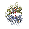
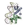
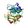
| Title | Salmonella enterica SafA pilin, head-to-tail swapped dimer of Ntd1 mutant | ||||||
 Components Components | SAFA PILUS SUBUNIT | ||||||
 Keywords Keywords |  FIBRIL PROTEIN / PILUS SUBUNIT / ADHESION / PATHOGENESIS / FOLD COMPLEMENTATION FIBRIL PROTEIN / PILUS SUBUNIT / ADHESION / PATHOGENESIS / FOLD COMPLEMENTATION | ||||||
| Function / homology |  Function and homology information Function and homology informationSaf-pilin pilus formation protein / Saf-pilin pilus formation protein / Dr adhesin / Dr-adhesin superfamily / Adhesion domain superfamily / Prokaryotic membrane lipoprotein lipid attachment site profile. / Immunoglobulin-like / Sandwich / Mainly BetaSimilarity search - Domain/homology | ||||||
| Biological species |  SALMONELLA TYPHIMURIUM (bacteria) SALMONELLA TYPHIMURIUM (bacteria) | ||||||
| Method | X-RAY DIFFRACTION / SAD / Resolution: 1.78 Å | ||||||
 Authors Authors | Remaut, H. / Rose, R.J. / Hannan, T.J. / Hultgren, S.J. / Radford, S.E. / Ashcroft, A.E. / Waksman, G. | ||||||
 Citation Citation | Journal: Mol.Cell / Year: 2006 Title: Donor-Strand Exchange in Chaperone-Assisted Pilus Assembly Proceeds Through a Concerted Beta-Strand Displacement Mechanism Authors: Remaut, H. / Rose, R.J. / Hannan, T.J. / Hultgren, S.J. / Radford, S.E. / Ashcroft, A.E. / Waksman, G. | ||||||
| History |
|
- Structure visualization
| Structure viewer | Molecule: MolmilJmol/JSmol |
|---|
- Downloads & links
-Download
| PDBx/mmCIF format | 2co3.cif.gz | 67.8 KB | Display | PDBx/mmCIF format |
|---|---|---|---|---|
| PDB format | pdb2co3.ent.gz | 51.2 KB | Display | PDB format |
| PDBx/mmJSON format | 2co3.json.gz | Tree view | PDBx/mmJSON format | |
| Others |  Other downloads Other downloads |
-Validation report
| Arichive directory | https://data.pdbj.org/pub/pdb/validation_reports/co/2co3ftp://data.pdbj.org/pub/pdb/validation_reports/co/2co3 | HTTPS FTP |
|---|
-Related structure data
| Related structure data | 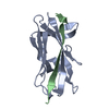 2cnyC  2cnzC 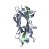 2co1C  2co2C 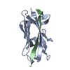 2co4C  2co6C 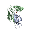 2co7C C: citing same article ( |
|---|---|
| Similar structure data |


-Links
 PDBj
PDBj


- Assembly
| Deposited unit | 
| ||||||||
|---|---|---|---|---|---|---|---|---|---|
| 1 |
| ||||||||
| Unit cell |
| ||||||||
| Details | FOR THE HETERO-ASSEMBLY DESCRIBED BY REMARK 350THE N-TERMINAL EXTENSION PEPTIDE OF ONE SUBUNIT, CHAIN B,INSERTS INTO THE FOLD OF ANOTHER, CHAIN A. THIS INTERACTIONIS REPETED IN THE POLYMER, AN N-TERMINAL EXTENSION OFMOLECULE C WOULD INSERT IN TO THE SUBUNIT OF MOLECULE B , DINTO C ETC |
-Components
| #1: Protein | Mass: 14454.134 Da / Num. of mol.: 2 / Fragment: RESIDUES 27-28 AND 36-170 Source method: isolated from a genetically manipulated source Source: (gene. exp.) SALMONELLA TYPHIMURIUM (bacteria) / Strain: LT2 / Plasmid: PTRC99A / Production host: ESCHERICHIA COLI (E. coli) / Strain (production host): C600 / References: UniProt: Q8ZRK4#2: Water | ChemComp-HOH / | Water Mass: 18.015 Da / Num. of mol.: 257 / Source method: isolated from a natural source / Formula: H2O Mass: 18.015 Da / Num. of mol.: 257 / Source method: isolated from a natural source / Formula: H2OSequence details | RESIDUES 3-9 WERE DELETED IN THE SAMPLE USED IN THE EXPERIMENT | |
|---|
-Experimental details
-Experiment
| Experiment | Method: X-RAY DIFFRACTION / Number of used crystals: 1 |
|---|
- Sample preparation
| Crystal | Density Matthews: 1.84 Å3/Da / Density % sol: 32.5 % |
|---|---|
| Crystal grow | pH: 5.6 Details: 30% PEG 4000, 100 MM MES, PH 5.6, 200 MM AMMONIUM ACETATE |
-Data collection
| Diffraction | Mean temperature: 100 K |
|---|---|
| Diffraction source | Source: ROTATING ANODE / Type: RIGAKU R-AXIS / Wavelength: 1.5418 |
| Detector | Type: MARRESEARCH / Detector: IMAGE PLATE |
| Radiation | Protocol: SINGLE WAVELENGTH / Monochromatic (M) / Laue (L): M / Scattering type: x-ray |
| Radiation wavelength | Wavelength: 1.5418 Å / Relative weight: 1 |
| Reflection | Resolution: 1.78→20 Å / Num. obs: 201192 / % possible obs: 97.3 % / Observed criterion σ(I): 0 / Redundancy: 2.9 % / Biso Wilson estimate: 15.8 Å2 / Rmerge(I) obs: 0.05 / Net I/σ(I): 22.8 |
| Reflection shell | Resolution: 1.78→1.84 Å / Redundancy: 3 % / Rmerge(I) obs: 0.18 / Mean I/σ(I) obs: 5.1 / % possible all: 95.7 |
- Processing
| Software |
| ||||||||||||||||||||||||||||||||||||||||||||||||||||||||||||
|---|---|---|---|---|---|---|---|---|---|---|---|---|---|---|---|---|---|---|---|---|---|---|---|---|---|---|---|---|---|---|---|---|---|---|---|---|---|---|---|---|---|---|---|---|---|---|---|---|---|---|---|---|---|---|---|---|---|---|---|---|---|
| Refinement | Method to determine structure: SAD / Resolution: 1.78→15 Å / Data cutoff high absF: 10000 / Cross valid method: THROUGHOUT / σ(F): 0 Details: SIDE CHAINS WITH NO VISIBLE ELECTRON DENSITY ABOVE 1 SIGMA HAVE OCCUPANCY SET TO 0.05
| ||||||||||||||||||||||||||||||||||||||||||||||||||||||||||||
| Solvent computation | Bsol: 47.32 Å2 / ksol: 0.370467 e/Å3 | ||||||||||||||||||||||||||||||||||||||||||||||||||||||||||||
| Displacement parameters | Biso mean: 15.7 Å2
| ||||||||||||||||||||||||||||||||||||||||||||||||||||||||||||
| Refinement step | Cycle: LAST / Resolution: 1.78→15 Å
| ||||||||||||||||||||||||||||||||||||||||||||||||||||||||||||
| Refine LS restraints |
| ||||||||||||||||||||||||||||||||||||||||||||||||||||||||||||
| Xplor file |
|
