 Movie
Movie Controller


+ Open data

- Basic information
| Entry | Database: PDB / ID: 2cn5 | ||||||
|---|---|---|---|---|---|---|---|
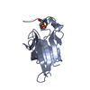
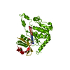
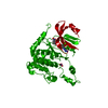
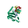
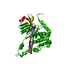
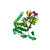
| Title | Crystal structure of human Chk2 in complex with ADP | ||||||
 Components Components | SERINE/THREONINE-PROTEIN KINASE CHK2 | ||||||
 Keywords Keywords |  TRANSFERASE / KINASE / KINASE DOMAIN / CHECKPOINT / CANCER / TUMOUR SUPPRESSOR / CHEK2 / CHK2 / CDS1 / RAD53 / PHOSPHORYLATION / ACTIVATION SEGMENT / LI-FRAUMENI SYNDROME / ATP-BINDING / CELL CYCLE / DISEASE MUTATION / MAGNESIUM / METAL-BINDING / NUCLEAR PROTEIN / NUCLEOTIDE-BINDING / PROTO-ONCOGENE / SERINE/THREONINE-PROTEIN KINASE TRANSFERASE / KINASE / KINASE DOMAIN / CHECKPOINT / CANCER / TUMOUR SUPPRESSOR / CHEK2 / CHK2 / CDS1 / RAD53 / PHOSPHORYLATION / ACTIVATION SEGMENT / LI-FRAUMENI SYNDROME / ATP-BINDING / CELL CYCLE / DISEASE MUTATION / MAGNESIUM / METAL-BINDING / NUCLEAR PROTEIN / NUCLEOTIDE-BINDING / PROTO-ONCOGENE / SERINE/THREONINE-PROTEIN KINASE | ||||||
| Function / homology |  Function and homology information Function and homology informationmitotic DNA damage checkpoint signaling / mitotic intra-S DNA damage checkpoint signaling / DNA damage response, signal transduction by p53 class mediator resulting in transcription of p21 class mediator / thymocyte apoptotic process / regulation of protein catabolic process / replicative senescence / mitotic spindle assembly / intrinsic apoptotic signaling pathway in response to DNA damage by p53 class mediator / signal transduction in response to DNA damage / Chk1/Chk2(Cds1) mediated inactivation of Cyclin B:Cdk1 complex ...mitotic DNA damage checkpoint signaling / mitotic intra-S DNA damage checkpoint signaling / DNA damage response, signal transduction by p53 class mediator resulting in transcription of p21 class mediator / thymocyte apoptotic process / regulation of protein catabolic process / replicative senescence / mitotic spindle assembly / intrinsic apoptotic signaling pathway in response to DNA damage by p53 class mediator / signal transduction in response to DNA damage / Chk1/Chk2(Cds1) mediated inactivation of Cyclin B:Cdk1 complex / DNA damage response, signal transduction by p53 class mediator resulting in cell cycle arrest / regulation of signal transduction by p53 class mediator / DNA damage checkpoint signaling / Ubiquitin Mediated Degradation of Phosphorylated Cdc25A / Stabilization of p53 / protein catabolic process / G2/M DNA damage checkpoint / Regulation of TP53 Activity through Methylation / cellular response to gamma radiation / PML body / G2/M transition of mitotic cell cycle / intrinsic apoptotic signaling pathway in response to DNA damage / double-strand break repair / Regulation of TP53 Degradation / Recruitment and ATM-mediated phosphorylation of repair and signaling proteins at DNA double strand breaks / Regulation of TP53 Activity through Phosphorylation / protein autophosphorylation / protein stabilization / non-specific serine/threonine protein kinase / cell division / protein phosphorylation / protein serine kinase activity / protein serine/threonine kinase activity / DNA damage response / ubiquitin protein ligase binding / regulation of DNA-templated transcription / protein kinase binding / positive regulation of DNA-templated transcription / Golgi apparatus / protein homodimerization activity / nucleoplasm / ATP binding / identical protein binding / metal ion binding / nucleus / cytoplasmSimilarity search - Function | ||||||
| Biological species |  HOMO SAPIENS (human) HOMO SAPIENS (human) | ||||||
| Method | X-RAY DIFFRACTION / SYNCHROTRON / MOLECULAR REPLACEMENT / Resolution: 2.25 Å | ||||||
 Authors Authors | Oliver, A.W. / Pearl, L.H. | ||||||
 Citation Citation | Journal: Embo J. / Year: 2006 Title: Trans-Activation of the DNA-Damage Signalling Protein Kinase Chk2 by T-Loop Exchange Authors: Oliver, A.W. / Paul, A. / Boxall, K.J. / Barrie, S.E. / Aherne, G.W. / Garrett, M.D. / Mittnacht, S. / Pearl, L.H. | ||||||
| History |
|
- Structure visualization
| Structure viewer | Molecule: MolmilJmol/JSmol |
|---|
- Downloads & links
-Download
| PDBx/mmCIF format | 2cn5.cif.gz | 78.8 KB | Display | PDBx/mmCIF format |
|---|---|---|---|---|
| PDB format | pdb2cn5.ent.gz | 56.8 KB | Display | PDB format |
| PDBx/mmJSON format | 2cn5.json.gz | Tree view | PDBx/mmJSON format | |
| Others |  Other downloads Other downloads |
-Validation report
| Arichive directory | https://data.pdbj.org/pub/pdb/validation_reports/cn/2cn5ftp://data.pdbj.org/pub/pdb/validation_reports/cn/2cn5 | HTTPS FTP |
|---|
-Related structure data
| Related structure data | 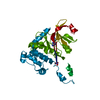 2cn8C 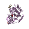 1atpS C: citing same article ( S: Starting model for refinement |
|---|---|
| Similar structure data |


-Links
 PDBj
PDBj


- Assembly
| Deposited unit | 
| ||||||||
|---|---|---|---|---|---|---|---|---|---|
| 1 | 
| ||||||||
| Unit cell |
|
-Components
-Protein , 1 types, 1 molecules A
| #1: Protein | Mass: 37111.844 Da / Num. of mol.: 1 / Fragment: KINASE DOMAIN, RESIDUES 210-531 Source method: isolated from a genetically manipulated source Source: (gene. exp.) HOMO SAPIENS (human) / Description: HUMAN CDNA LIBRARY / Plasmid: PTHREE-E / Production host:  ESCHERICHIA COLI (E. coli) / Strain (production host): ROSETTA2 (DE3) PLYSS ESCHERICHIA COLI (E. coli) / Strain (production host): ROSETTA2 (DE3) PLYSSReferences: UniProt: O96017, non-specific serine/threonine protein kinase |
|---|
-Non-polymers , 5 types, 159 molecules 


| #2: Chemical | ChemComp-CL / Chloride Mass: 35.453 Da / Num. of mol.: 1 / Source method: obtained synthetically / Formula: Cl Mass: 35.453 Da / Num. of mol.: 1 / Source method: obtained synthetically / Formula: Cl | ||||||
|---|---|---|---|---|---|---|---|
| #3: Chemical |  Mass: 24.305 Da / Num. of mol.: 2 / Source method: obtained synthetically / Formula: Mg Mass: 24.305 Da / Num. of mol.: 2 / Source method: obtained synthetically / Formula: Mg#4: Chemical | ChemComp-NO3 / | Nitrate Mass: 62.005 Da / Num. of mol.: 1 / Source method: obtained synthetically / Formula: NO3 Mass: 62.005 Da / Num. of mol.: 1 / Source method: obtained synthetically / Formula: NO3#5: Chemical | ChemComp-ADP / | Adenosine diphosphate Mass: 427.201 Da / Num. of mol.: 1 / Source method: obtained synthetically / Formula: C10H15N5O10P2 / Comment: ADP, energy-carrying molecule*YM Mass: 427.201 Da / Num. of mol.: 1 / Source method: obtained synthetically / Formula: C10H15N5O10P2 / Comment: ADP, energy-carrying molecule*YM#6: Water | ChemComp-HOH / | WaterMass: 18.015 Da / Num. of mol.: 154 / Source method: isolated from a natural source / Formula: H2O |
-Experimental details
-Experiment
| Experiment | Method: X-RAY DIFFRACTION / Number of used crystals: 1 |
|---|
- Sample preparation
| Crystal | Density Matthews: 3.02 Å3/Da / Density % sol: 59.3 % |
|---|---|
| Crystal grow | Details: 0.1 M HEPES PH 7.5, 0.2 M MAGNESIUM NITRATE, 8-16% (W/V) PEG 3350 |
-Data collection
| Diffraction | Mean temperature: 100 K |
|---|---|
| Diffraction source | Source: SYNCHROTRON / Site: ESRF  / Beamline: ID29 / Wavelength: 0.9151 / Beamline: ID29 / Wavelength: 0.9151 |
| Detector | Type: ADSC CCD / Detector: CCD / Date: Sep 7, 2005 |
| Radiation | Protocol: SINGLE WAVELENGTH / Monochromatic (M) / Laue (L): M / Scattering type: x-ray |
| Radiation wavelength | Wavelength: 0.9151 Å / Relative weight: 1 |
| Reflection | Resolution: 2.25→40.82 Å / Num. obs: 21525 / % possible obs: 100 % / Observed criterion σ(I): 2 / Redundancy: 5.5 % / Rmerge(I) obs: 0.08 / Net I/σ(I): 16.5 |
| Reflection shell | Resolution: 2.25→2.37 Å / Redundancy: 5.6 % / Rmerge(I) obs: 0.47 / Mean I/σ(I) obs: 3.2 / % possible all: 100 |
- Processing
| Software |
| ||||||||||||||||||||||||||||||||||||||||||||||||||||||||||||||||||||||||||||||||||||||||||||||||||||||||||||||||||||||||||||||||||||||||||||||||||||||||||||||||||||||||||||||||||||||
|---|---|---|---|---|---|---|---|---|---|---|---|---|---|---|---|---|---|---|---|---|---|---|---|---|---|---|---|---|---|---|---|---|---|---|---|---|---|---|---|---|---|---|---|---|---|---|---|---|---|---|---|---|---|---|---|---|---|---|---|---|---|---|---|---|---|---|---|---|---|---|---|---|---|---|---|---|---|---|---|---|---|---|---|---|---|---|---|---|---|---|---|---|---|---|---|---|---|---|---|---|---|---|---|---|---|---|---|---|---|---|---|---|---|---|---|---|---|---|---|---|---|---|---|---|---|---|---|---|---|---|---|---|---|---|---|---|---|---|---|---|---|---|---|---|---|---|---|---|---|---|---|---|---|---|---|---|---|---|---|---|---|---|---|---|---|---|---|---|---|---|---|---|---|---|---|---|---|---|---|---|---|---|---|
| Refinement | Method to determine structure: MOLECULAR REPLACEMENT Starting model: PDB ENTRY 1ATP Resolution: 2.25→40.83 Å / Cor.coef. Fo:Fc: 0.947 / Cor.coef. Fo:Fc free: 0.928 / SU B: 9.557 / SU ML: 0.131 / TLS residual ADP flag: LIKELY RESIDUAL / Cross valid method: THROUGHOUT / ESU R: 0.22 / ESU R Free: 0.191 / Stereochemistry target values: MAXIMUM LIKELIHOOD Details: THE WATER MOLECULES IN CHAIN Y TRACE THE PATH OF THE EXTREME C-TERMINUS OF THE PROTEIN. SIDE CHAIN IDENTITIES COULD NOT BE DETERMINED FOR MODELLING.
| ||||||||||||||||||||||||||||||||||||||||||||||||||||||||||||||||||||||||||||||||||||||||||||||||||||||||||||||||||||||||||||||||||||||||||||||||||||||||||||||||||||||||||||||||||||||
| Solvent computation | Ion probe radii: 0.8 Å / Shrinkage radii: 0.8 Å / VDW probe radii: 1.4 Å / Solvent model: MASK | ||||||||||||||||||||||||||||||||||||||||||||||||||||||||||||||||||||||||||||||||||||||||||||||||||||||||||||||||||||||||||||||||||||||||||||||||||||||||||||||||||||||||||||||||||||||
| Displacement parameters | Biso mean: 46.59 Å2
| ||||||||||||||||||||||||||||||||||||||||||||||||||||||||||||||||||||||||||||||||||||||||||||||||||||||||||||||||||||||||||||||||||||||||||||||||||||||||||||||||||||||||||||||||||||||
| Refinement step | Cycle: LAST / Resolution: 2.25→40.83 Å
| ||||||||||||||||||||||||||||||||||||||||||||||||||||||||||||||||||||||||||||||||||||||||||||||||||||||||||||||||||||||||||||||||||||||||||||||||||||||||||||||||||||||||||||||||||||||
| Refine LS restraints |
|