- PDB-2clb: The structure of the DPS-like protein from Sulfolobus solfataricu... -
+
Open data
ID or keywords:
Loading...
-
Basic information
Entry
Database: PDB / ID: 2clb
Title
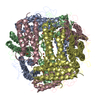
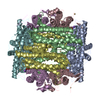
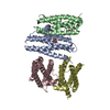
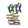
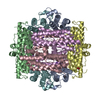
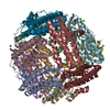
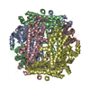
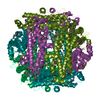
The structure of the DPS-like protein from Sulfolobus solfataricus reveals a bacterioferritin-like di-metal binding site within a Dps- like dodecameric assembly
Components
DPS-LIKE PROTEIN
Keywords
METAL BINDING PROTEIN / DI-IRON CARBOXYLATE / HYPOTHETICAL PROTEIN / BACTERIOFERRITIN / HYDROGEN PEROXIDE / DPS / ARCHAEA / DPS- LIKE / OXIDATIVE STRESS
Function / homology
Function and homology information
Oxidoreductases; Oxidizing metal ions / nucleoid / intracellular sequestering of iron ion / ferroxidase activity / ferric iron binding / iron ion binding / heme binding / cytosol Similarity search - Function
A: DPS-LIKE PROTEIN B: DPS-LIKE PROTEIN C: DPS-LIKE PROTEIN D: DPS-LIKE PROTEIN M: DPS-LIKE PROTEIN N: DPS-LIKE PROTEIN O: DPS-LIKE PROTEIN P: DPS-LIKE PROTEIN hetero molecules
 Movie
Movie Controller

 Yorodumi
 Open data
 Basic information
 Components
 Keywords
 HYPOTHETICAL PROTEIN /
  Function and homology information

 Authors
 Citation
 Structure visualization
 Downloads & links
 Other downloads
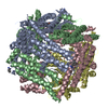

 PDBj
PDBj


 Assembly


 Mass: 65.409 Da / Num. of mol.: 8 / Source method: obtained synthetically / Formula: Zn
 Mass: 55.845 Da / Num. of mol.: 8 / Source method: obtained synthetically / Formula: Fe
 Mass: 18.015 Da / Num. of mol.: 121 / Source method: isolated from a natural source / Formula: H2O
 Sample preparation
 / Beamline: BL9-2 / Wavelength: 1.28237
  Processing