 Movie
Movie Controller

[English] 日本語
 Yorodumi
- PDB-2cks: X-RAY CRYSTAL STRUCTURE OF THE CATALYTIC DOMAIN OF THERMOBIFIDA F... -
+ Open data

- Basic information
| Entry | Database: PDB / ID: 2cks | |||||||||
|---|---|---|---|---|---|---|---|---|---|---|
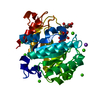
| Title | X-RAY CRYSTAL STRUCTURE OF THE CATALYTIC DOMAIN OF THERMOBIFIDA FUSCA ENDOGLUCANASE CEL5A (E5) | |||||||||
 Components Components | ENDOGLUCANASE E-5 | |||||||||
 Keywords Keywords |  HYDROLASE / CARBOHYDRATE METABOLISM / POLYSACCHARIDE DEGRADATION / GLYCOSIDE HYDROLASE FAMILY 5 / GLYCOSIDASE / ENDOGLUCANASE / THERMOBIFIDA FUSCA E5 / CELLULOSE DEGRADATION HYDROLASE / CARBOHYDRATE METABOLISM / POLYSACCHARIDE DEGRADATION / GLYCOSIDE HYDROLASE FAMILY 5 / GLYCOSIDASE / ENDOGLUCANASE / THERMOBIFIDA FUSCA E5 / CELLULOSE DEGRADATION | |||||||||
| Function / homology |  Function and homology informationcellulase / polysaccharide binding / cellulase activity / cellulose catabolic process Function and homology informationcellulase / polysaccharide binding / cellulase activity / cellulose catabolic processSimilarity search - Function | |||||||||
| Biological species |   THERMOBIFIDA FUSCA (bacteria) THERMOBIFIDA FUSCA (bacteria) | |||||||||
| Method | X-RAY DIFFRACTION / SYNCHROTRON / MOLECULAR REPLACEMENT / Resolution: 1.6 Å | |||||||||
 Authors Authors | Berglund, G.I. / Gualfetti, P.J. / Requadt, C. / Gross, L.S. / Bergfors, T. / Shaw, A. / Saldajeno, M. / Mitchinson, C. / Sandgren, M. | |||||||||
 Citation Citation | Journal: To be Published Title: The Crystal Structure of the Catalytic Domain of Thermobifida Fusca Endoglucanase Cel5A in Complex with Cellotetraose Authors: Berglund, G.I. / Gualfetti, P.J. / Requadt, C. / Gross, L.S. / Bergfors, T. / Shaw, A. / Saldajeno, M. / Mitchinson, C. / Sandgren, M. | |||||||||
| History |
| |||||||||
| Remark 700 | SHEET DETERMINATION METHOD: DSSP THE SHEETS PRESENTED AS "AB" IN EACH CHAIN ON SHEET RECORDS BELOW ... SHEET DETERMINATION METHOD: DSSP THE SHEETS PRESENTED AS "AB" IN EACH CHAIN ON SHEET RECORDS BELOW IS ACTUALLY AN 8-STRANDED BARREL THIS IS REPRESENTED BY A 9-STRANDED SHEET IN WHICH THE FIRST AND LAST STRANDS ARE IDENTICAL. THE SHEETS PRESENTED AS "BB" IN EACH CHAIN ON SHEET RECORDS BELOW IS ACTUALLY AN 8-STRANDED BARREL THIS IS REPRESENTED BY A 9-STRANDED SHEET IN WHICH THE FIRST AND LAST STRANDS ARE IDENTICAL. |
- Structure visualization
| Structure viewer | Molecule: MolmilJmol/JSmol |
|---|
- Downloads & links
-Download
| PDBx/mmCIF format | 2cks.cif.gz | 156.2 KB | Display | PDBx/mmCIF format |
|---|---|---|---|---|
| PDB format | pdb2cks.ent.gz | 122.5 KB | Display | PDB format |
| PDBx/mmJSON format | 2cks.json.gz | Tree view | PDBx/mmJSON format | |
| Others |  Other downloads Other downloads |
-Validation report
| Arichive directory | https://data.pdbj.org/pub/pdb/validation_reports/ck/2cksftp://data.pdbj.org/pub/pdb/validation_reports/ck/2cks | HTTPS FTP |
|---|
-Related structure data


-Links
 PDBj
PDBj

- Assembly
| Deposited unit | 
| ||||||||
|---|---|---|---|---|---|---|---|---|---|
| 1 | 
| ||||||||
| 2 | 
| ||||||||
| Unit cell |
|
-Components
| #1: Protein | Mass: 34176.965 Da / Num. of mol.: 2 / Fragment: CATALYTIC DOMAIN, RESIDUES 161-466 Source method: isolated from a genetically manipulated source Source: (gene. exp.) THERMOBIFIDA FUSCA (bacteria) / Plasmid: PBS42 / Production host: BACILLUS SUBTILIS (bacteria) / References: UniProt: Q01786, cellulase#2: Chemical | Benzamidine  Mass: 120.152 Da / Num. of mol.: 2 / Source method: obtained synthetically / Formula: C7H8N2 Mass: 120.152 Da / Num. of mol.: 2 / Source method: obtained synthetically / Formula: C7H8N2#3: Chemical | ChemComp-ZN /   Mass: 65.409 Da / Num. of mol.: 8 / Source method: obtained synthetically / Formula: Zn Mass: 65.409 Da / Num. of mol.: 8 / Source method: obtained synthetically / Formula: Zn#4: Chemical | ChemComp-NA /   Mass: 22.990 Da / Num. of mol.: 4 / Source method: obtained synthetically / Formula: Na Mass: 22.990 Da / Num. of mol.: 4 / Source method: obtained synthetically / Formula: Na#5: Water | ChemComp-HOH / | Water Mass: 18.015 Da / Num. of mol.: 640 / Source method: isolated from a natural source / Formula: H2O Mass: 18.015 Da / Num. of mol.: 640 / Source method: isolated from a natural source / Formula: H2OCompound details | ENDOHYDROL | |
|---|
-Experimental details
-Experiment
| Experiment | Method: X-RAY DIFFRACTION / Number of used crystals: 1 |
|---|
- Sample preparation
| Crystal | Density Matthews: 1.8 Å3/Da / Density % sol: 30.3 % |
|---|---|
| Crystal grow | Method: vapor diffusion, hanging drop / pH: 5.2 Details: PROTEIN WAS CRYSTALLISED USING THE HANGING DROP VAPOUR DIFFUSION METHOD BY MIXING 2 MICROLITER OF WELL SOLUTION CONTAINING 10 % PEG 8000, 0.1 M HEPES PH 7.0, 0.1 M NA ACETATE, 5MM ZN ACETATE ...Details: PROTEIN WAS CRYSTALLISED USING THE HANGING DROP VAPOUR DIFFUSION METHOD BY MIXING 2 MICROLITER OF WELL SOLUTION CONTAINING 10 % PEG 8000, 0.1 M HEPES PH 7.0, 0.1 M NA ACETATE, 5MM ZN ACETATE WITH 2 MICROLITER 1MG/ML PROTEIN SOLUTION AND 0.5 MICROLITER OF 20 % BENZAMIDINE |
-Data collection
| Diffraction | Mean temperature: 100 K |
|---|---|
| Diffraction source | Source: SYNCHROTRON / Site: ESRF  / Beamline: ID14-2 / Wavelength: 0.933 / Beamline: ID14-2 / Wavelength: 0.933 |
| Detector | Type: ADSC CCD / Detector: CCD / Date: Sep 28, 2002 |
| Radiation | Protocol: SINGLE WAVELENGTH / Monochromatic (M) / Laue (L): M / Scattering type: x-ray |
| Radiation wavelength | Wavelength: 0.933 Å / Relative weight: 1 |
| Reflection | Resolution: 1.6→42.3 Å / Num. obs: 67888 / % possible obs: 99.8 % / Redundancy: 4 % / Rmerge(I) obs: 0.08 / Net I/σ(I): 15.1 |
| Reflection shell | Resolution: 1.6→1.64 Å / Redundancy: 3.4 % / Rmerge(I) obs: 0.27 / Mean I/σ(I) obs: 4.1 / % possible all: 99.8 |
- Processing
| Software |
| ||||||||||||||||||||||||||||||||||||||||||||||||||||||||||||||||||||||||||||||||||||||||||||||||||||||||||||||||||||||||||||||||||||||||||||||||||||||||||||||||||||||||||||||||||||||
|---|---|---|---|---|---|---|---|---|---|---|---|---|---|---|---|---|---|---|---|---|---|---|---|---|---|---|---|---|---|---|---|---|---|---|---|---|---|---|---|---|---|---|---|---|---|---|---|---|---|---|---|---|---|---|---|---|---|---|---|---|---|---|---|---|---|---|---|---|---|---|---|---|---|---|---|---|---|---|---|---|---|---|---|---|---|---|---|---|---|---|---|---|---|---|---|---|---|---|---|---|---|---|---|---|---|---|---|---|---|---|---|---|---|---|---|---|---|---|---|---|---|---|---|---|---|---|---|---|---|---|---|---|---|---|---|---|---|---|---|---|---|---|---|---|---|---|---|---|---|---|---|---|---|---|---|---|---|---|---|---|---|---|---|---|---|---|---|---|---|---|---|---|---|---|---|---|---|---|---|---|---|---|---|
| Refinement | Method to determine structure: MOLECULAR REPLACEMENT Starting model: THEORETICAL MODEL PRODUCED BY SWISS-MODEL Resolution: 1.6→42.26 Å / Cor.coef. Fo:Fc: 0.969 / Cor.coef. Fo:Fc free: 0.955 / Cross valid method: THROUGHOUT / ESU R: 0.087 / ESU R Free: 0.083 / Stereochemistry target values: MAXIMUM LIKELIHOOD Details: HYDROGENS HAVE BEEN ADDED IN THE RIDING POSITIONS. THE FOLLOWING RESIDUES HAVE BEEN MODELLED IN MULTIPLE CONFORMATIONS A136, A138, A143, A166, A169, A206, A207, A248, A263, A271, A274, A289, ...Details: HYDROGENS HAVE BEEN ADDED IN THE RIDING POSITIONS. THE FOLLOWING RESIDUES HAVE BEEN MODELLED IN MULTIPLE CONFORMATIONS A136, A138, A143, A166, A169, A206, A207, A248, A263, A271, A274, A289, A290, A305, A323, A337, A343, A378, A388, A416, A425, A1431, B131, B138, B143, B166, B169, B206, B225, B248, B249, B252, B263, B271, B274, B289, B290, B297, B321, B343, B422, B1431, Y46, Y69, Y71, Y82, Y128, Y172, Y215, Y221, Y260, Y267, Z137, Z187, Z246, Z256, Z273, Z277, Z279, Z284, Z289. ATOMS WITH MISSING ELECTRON DENSITY ARE ASSIGNED ZERO OCCUPANCY. ATOMS ARE ASSIGNED REDUCED OCCUPANCIES WHEN ELECTRON DENSITY IS WEAK OR ATOMS HAVE PARTIAL OCCUPANCY. NO NCS CONSTRAINTS OR RESTRAINTS HAVE BEEN USED IN REFINEMENT OF THE TWO PROTEIN CHAINS IN THE ASYMMETRIC UNIT.
| ||||||||||||||||||||||||||||||||||||||||||||||||||||||||||||||||||||||||||||||||||||||||||||||||||||||||||||||||||||||||||||||||||||||||||||||||||||||||||||||||||||||||||||||||||||||
| Solvent computation | Ion probe radii: 0.8 Å / Shrinkage radii: 0.8 Å / VDW probe radii: 1.4 Å / Solvent model: BABINET MODEL PLUS MASK | ||||||||||||||||||||||||||||||||||||||||||||||||||||||||||||||||||||||||||||||||||||||||||||||||||||||||||||||||||||||||||||||||||||||||||||||||||||||||||||||||||||||||||||||||||||||
| Displacement parameters | Biso mean: 8.97 Å2
| ||||||||||||||||||||||||||||||||||||||||||||||||||||||||||||||||||||||||||||||||||||||||||||||||||||||||||||||||||||||||||||||||||||||||||||||||||||||||||||||||||||||||||||||||||||||
| Refinement step | Cycle: LAST / Resolution: 1.6→42.26 Å
| ||||||||||||||||||||||||||||||||||||||||||||||||||||||||||||||||||||||||||||||||||||||||||||||||||||||||||||||||||||||||||||||||||||||||||||||||||||||||||||||||||||||||||||||||||||||
| Refine LS restraints |
|