 Movie
Movie Controller

[English] 日本語
 Yorodumi
- PDB-2cjj: Crystal Structure of the MYB domain of the RAD transcription fact... -
+ Open data

- Basic information
| Entry | Database: PDB / ID: 2cjj | ||||||
|---|---|---|---|---|---|---|---|
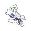
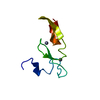
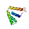
| Title | Crystal Structure of the MYB domain of the RAD transcription factor from Antirrhinum majus | ||||||
 Components Components | RADIALIS | ||||||
 Keywords Keywords |  DNA BINDING PROTEIN / PLANT DEVELOPMENT / DNA-BINDING PROTEIN / MYB TRANSCRIPTION FACTOR / DNA-BINDING / NUCLEAR PROTEIN / FLORAL ASYMMETRY DNA BINDING PROTEIN / PLANT DEVELOPMENT / DNA-BINDING PROTEIN / MYB TRANSCRIPTION FACTOR / DNA-BINDING / NUCLEAR PROTEIN / FLORAL ASYMMETRY | ||||||
| Function / homology |  Function and homology information Function and homology informationdetermination of dorsal/ventral asymmetry / flower development / nucleusSimilarity search - Function | ||||||
| Biological species |  ANTIRRHINUM MAJUS (snapdragon) ANTIRRHINUM MAJUS (snapdragon) | ||||||
| Method | X-RAY DIFFRACTION / SIRAS / Resolution: 1.9 Å | ||||||
 Authors Authors | Stevenson, C.E.M. / Burton, N. / Costa, M.M. / Nath, U. / Dixon, R.A. / Coen, E.S. / Lawson, D.M. | ||||||
 Citation Citation | Journal: Proteins: Struct., Funct., Bioinf. / Year: 2006 Title: Crystal Structure of the Myb Domain of the Rad Transcription Factor from Antirrhinum Majus. Authors: Stevenson, C.E.M. / Burton, N. / Costa, M.M. / Nath, U. / Dixon, R.A. / Coen, E.S. / Lawson, D.M. #1: Journal: Acta Crystallogr.,Sect.F / Year: 2005 Title: Crystallisation and Preliminary X-Ray Analysis of the Rad Protein from Antirrhinum Majus Authors: Stevenson, C.E.M. / Burton, N. / Costa, M. / Nath, U. / Dixon, R.A. / Coen, E.S. / Lawson, D.M. | ||||||
| History |
|
- Structure visualization
| Structure viewer | Molecule: MolmilJmol/JSmol |
|---|
- Downloads & links
-Download
| PDBx/mmCIF format | 2cjj.cif.gz | 26 KB | Display | PDBx/mmCIF format |
|---|---|---|---|---|
| PDB format | pdb2cjj.ent.gz | 17 KB | Display | PDB format |
| PDBx/mmJSON format | 2cjj.json.gz | Tree view | PDBx/mmJSON format | |
| Others |  Other downloads Other downloads |
-Validation report
| Arichive directory | https://data.pdbj.org/pub/pdb/validation_reports/cj/2cjjftp://data.pdbj.org/pub/pdb/validation_reports/cj/2cjj | HTTPS FTP |
|---|
-Related structure data
| Similar structure data |
|---|


-Links
 PDBj
PDBj
- Assembly
| Deposited unit | 
| ||||||||
|---|---|---|---|---|---|---|---|---|---|
| 1 |
| ||||||||
| Unit cell |
|
-Components
| #1: Protein | Mass: 10795.235 Da / Num. of mol.: 1 Source method: isolated from a genetically manipulated source Details: 8 KDA PROTEOLYTIC FRAGMENT, MYB DOMAIN / Source: (gene. exp.) ANTIRRHINUM MAJUS (snapdragon) / Plasmid: PRSET A / Production host:  ESCHERICHIA COLI (E. coli) / Strain (production host): BL21(DE3) / References: UniProt: Q58FS3 ESCHERICHIA COLI (E. coli) / Strain (production host): BL21(DE3) / References: UniProt: Q58FS3 |
|---|---|
| #2: Water | ChemComp-HOH / Water Mass: 18.015 Da / Num. of mol.: 49 / Source method: isolated from a natural source / Formula: H2O Mass: 18.015 Da / Num. of mol.: 49 / Source method: isolated from a natural source / Formula: H2O |
-Experimental details
-Experiment
| Experiment | Method: X-RAY DIFFRACTION / Number of used crystals: 1 |
|---|
- Sample preparation
| Crystal | Density Matthews: 2.3 Å3/Da / Density % sol: 46 % |
|---|---|
| Crystal grow | pH: 9.5 / Details: pH 9.50 |
-Data collection
| Diffraction | Mean temperature: 100 K |
|---|---|
| Diffraction source | Source: ROTATING ANODE / Type: RIGAKU RUH3R / Wavelength: 1.5418 |
| Detector | Type: MARRESEARCH / Detector: IMAGE PLATE / Date: May 18, 2004 / Details: MIRRORS |
| Radiation | Protocol: SINGLE WAVELENGTH / Monochromatic (M) / Laue (L): M / Scattering type: x-ray |
| Radiation wavelength | Wavelength: 1.5418 Å / Relative weight: 1 |
| Reflection | Resolution: 1.9→37.9 Å / Num. obs: 6112 / % possible obs: 98.6 % / Observed criterion σ(I): 3 / Redundancy: 10.8 % / Biso Wilson estimate: 26.1 Å2 / Rmerge(I) obs: 0.07 / Net I/σ(I): 33.3 |
| Reflection shell | Resolution: 1.9→1.97 Å / Rmerge(I) obs: 0.56 / Mean I/σ(I) obs: 3.6 / % possible all: 99.3 |
- Processing
| Software |
| ||||||||||||||||||||||||||||||||||||||||||||||||||||||||||||||||||||||||||||||||||||||||||||||||||||||||||||||||||||||||||||||||||||||||||||||||||||||||||||||||||||||||||||||||||||||
|---|---|---|---|---|---|---|---|---|---|---|---|---|---|---|---|---|---|---|---|---|---|---|---|---|---|---|---|---|---|---|---|---|---|---|---|---|---|---|---|---|---|---|---|---|---|---|---|---|---|---|---|---|---|---|---|---|---|---|---|---|---|---|---|---|---|---|---|---|---|---|---|---|---|---|---|---|---|---|---|---|---|---|---|---|---|---|---|---|---|---|---|---|---|---|---|---|---|---|---|---|---|---|---|---|---|---|---|---|---|---|---|---|---|---|---|---|---|---|---|---|---|---|---|---|---|---|---|---|---|---|---|---|---|---|---|---|---|---|---|---|---|---|---|---|---|---|---|---|---|---|---|---|---|---|---|---|---|---|---|---|---|---|---|---|---|---|---|---|---|---|---|---|---|---|---|---|---|---|---|---|---|---|---|
| Refinement | Method to determine structure: SIRAS / Resolution: 1.9→37.9 Å / Cor.coef. Fo:Fc: 0.949 / Cor.coef. Fo:Fc free: 0.93 / SU B: 9.067 / SU ML: 0.125 / TLS residual ADP flag: LIKELY RESIDUAL / Cross valid method: THROUGHOUT / ESU R: 0.168 / ESU R Free: 0.152 / Stereochemistry target values: MAXIMUM LIKELIHOOD / Details: HYDROGENS HAVE BEEN ADDED IN THE RIDING POSITIONS.
| ||||||||||||||||||||||||||||||||||||||||||||||||||||||||||||||||||||||||||||||||||||||||||||||||||||||||||||||||||||||||||||||||||||||||||||||||||||||||||||||||||||||||||||||||||||||
| Solvent computation | Ion probe radii: 0.8 Å / Shrinkage radii: 0.8 Å / VDW probe radii: 1.4 Å / Solvent model: MASK | ||||||||||||||||||||||||||||||||||||||||||||||||||||||||||||||||||||||||||||||||||||||||||||||||||||||||||||||||||||||||||||||||||||||||||||||||||||||||||||||||||||||||||||||||||||||
| Displacement parameters | Biso mean: 24.04 Å2
| ||||||||||||||||||||||||||||||||||||||||||||||||||||||||||||||||||||||||||||||||||||||||||||||||||||||||||||||||||||||||||||||||||||||||||||||||||||||||||||||||||||||||||||||||||||||
| Refinement step | Cycle: LAST / Resolution: 1.9→37.9 Å
| ||||||||||||||||||||||||||||||||||||||||||||||||||||||||||||||||||||||||||||||||||||||||||||||||||||||||||||||||||||||||||||||||||||||||||||||||||||||||||||||||||||||||||||||||||||||
| Refine LS restraints |
|