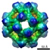
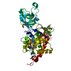
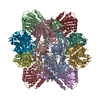
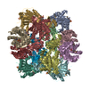
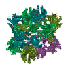
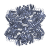
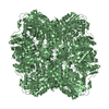
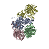
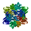
Journal: J Biol Chem / Year: 2006 Title: An archaeal peptidase assembles into two different quaternary structures: A tetrahedron and a giant octahedron. Authors: Guy Schoehn / Frédéric M D Vellieux / M Asunción Durá / Véronique Receveur-Bréchot / Céline M S Fabry / Rob W H Ruigrok / Christine Ebel / Alain Roussel / Bruno Franzetti / Abstract: Cellular proteolysis involves large oligomeric peptidases that play key roles in the regulation of many cellular processes. The cobalt-activated peptidase TET1 from the hyperthermophilic Archaea ...Cellular proteolysis involves large oligomeric peptidases that play key roles in the regulation of many cellular processes. The cobalt-activated peptidase TET1 from the hyperthermophilic Archaea Pyrococcus horikoshii (PhTET1) was found to assemble as a 12-subunit tetrahedron and as a 24-subunit octahedral particle. Both quaternary structures were solved by combining x-ray crystallography and cryoelectron microscopy data. The internal organization of the PhTET1 particles reveals highly self-compartmentalized systems made of networks of access channels extended by vast catalytic chambers. The two edifices display aminopeptidase activity, and their organizations indicate substrate navigation mechanisms different from those described in other large peptidase complexes. Compared with the tetrahedron, the octahedron forms a more expanded hollow structure, representing a new type of giant peptidase complex. PhTET1 assembles into two different quaternary structures because of quasi-equivalent contacts that previously have only been identified in viral capsids.
SHEET THE SHEET STRUCTURE OF THIS MOLECULE IS BIFURCATED. IN ORDER TO REPRESENT THIS FEATURE IN ... SHEET THE SHEET STRUCTURE OF THIS MOLECULE IS BIFURCATED. IN ORDER TO REPRESENT THIS FEATURE IN THE SHEET RECORDS BELOW, TWO SHEETS ARE DEFINED.
In the structure databanks used in Yorodumi, some data are registered as the other names, "COVID-19 virus" and "2019-nCoV". Here are the details of the virus and the list of structure data.
Jan 31, 2019. EMDB accession codes are about to change! (news from PDBe EMDB page)
EMDB accession codes are about to change! (news from PDBe EMDB page)
The allocation of 4 digits for EMDB accession codes will soon come to an end. Whilst these codes will remain in use, new EMDB accession codes will include an additional digit and will expand incrementally as the available range of codes is exhausted. The current 4-digit format prefixed with “EMD-” (i.e. EMD-XXXX) will advance to a 5-digit format (i.e. EMD-XXXXX), and so on. It is currently estimated that the 4-digit codes will be depleted around Spring 2019, at which point the 5-digit format will come into force.
The EM Navigator/Yorodumi systems omit the EMD- prefix.
Related info.:Q: What is EMD? / ID/Accession-code notation in Yorodumi/EM Navigator
Yorodumi is a browser for structure data from EMDB, PDB, SASBDB, etc.
This page is also the successor to EM Navigator detail page, and also detail information page/front-end page for Omokage search.
The word "yorodu" (or yorozu) is an old Japanese word meaning "ten thousand". "mi" (miru) is to see.
Related info.:EMDB / PDB / SASBDB / Comparison of 3 databanks / Yorodumi Search / Aug 31, 2016. New EM Navigator & Yorodumi / Yorodumi Papers / Jmol/JSmol / Function and homology information / Changes in new EM Navigator and Yorodumi
 Movie
Movie Controller

 Yorodumi
 Open data
 Basic information
 Components
 Keywords
 HYDROLASE /
  Function and homology information

 Authors
 Citation
 Structure visualization
 Downloads & links
 Other downloads
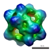
 PDBj
PDBj
 Assembly


 Mass: 58.933 Da / Num. of mol.: 2 / Source method: obtained synthetically / Formula: Co
 Sample preparation
 Processing