 Movie
Movie Controller

[English] 日本語
 Yorodumi
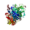
- PDB-2cer: Beta-glycosidase from Sulfolobus solfataricus in complex with phe... -
+ Open data

- Basic information
| Entry | Database: PDB / ID: 2cer | ||||||
|---|---|---|---|---|---|---|---|
| Title | Beta-glycosidase from Sulfolobus solfataricus in complex with phenethyl-substituted glucoimidazole | ||||||
 Components Components | BETA-GLUCOSIDASE A | ||||||
 Keywords Keywords |  HYDROLASE / GLYCOSIDE HYDROLASE / INHIBITOR / TRANSITION STATE MIMIC / FAMILY 1 / GLUCOIMIDAZOLE HYDROLASE / GLYCOSIDE HYDROLASE / INHIBITOR / TRANSITION STATE MIMIC / FAMILY 1 / GLUCOIMIDAZOLE | ||||||
| Function / homology |  Function and homology informationbeta-galactosidase / carbohydrate catabolic process / beta-galactosidase activity / beta-glucosidase activity / cytosol Function and homology informationbeta-galactosidase / carbohydrate catabolic process / beta-galactosidase activity / beta-glucosidase activity / cytosolSimilarity search - Function | ||||||
| Biological species |   SULFOLOBUS SOLFATARICUS (archaea) SULFOLOBUS SOLFATARICUS (archaea) | ||||||
| Method | X-RAY DIFFRACTION / SYNCHROTRON / MOLECULAR REPLACEMENT / Resolution: 2.29 Å | ||||||
 Authors Authors | Gloster, T.M. / Roberts, S. / Moracci, M. / Vasella, A. / Davies, G.J. | ||||||
 Citation Citation | Journal: Biochemistry / Year: 2006 Title: Structural, Kinetic, and Thermodynamic Analysis of Glucoimidazole-Derived Glycosidase Inhibitors. Authors: Gloster, T.M. / Roberts, S. / Perugino, G. / Rossi, M. / Moracci, M. / Panday, N. / Terinek, M. / Vasella, A. / Davies, G.J. | ||||||
| History |
| ||||||
| Remark 700 | SHEET DETERMINATION METHOD: DSSP THE SHEETS PRESENTED AS "AB" AND "BB" IN EACH CHAIN ON SHEET ... SHEET DETERMINATION METHOD: DSSP THE SHEETS PRESENTED AS "AB" AND "BB" IN EACH CHAIN ON SHEET RECORDS BELOW ARE ACTUALLY 9-STRANDED BARRELS THAT ARE REPRESENTED BY A 10-STRANDED SHEET IN WHICH THE FIRST AND LAST STRANDS ARE IDENTICAL. |
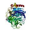
- Structure visualization
| Structure viewer | Molecule: MolmilJmol/JSmol |
|---|
- Downloads & links
-Download
| PDBx/mmCIF format | 2cer.cif.gz | 227.8 KB | Display | PDBx/mmCIF format |
|---|---|---|---|---|
| PDB format | pdb2cer.ent.gz | 185 KB | Display | PDB format |
| PDBx/mmJSON format | 2cer.json.gz | Tree view | PDBx/mmJSON format | |
| Others |  Other downloads Other downloads |
-Validation report
| Arichive directory | https://data.pdbj.org/pub/pdb/validation_reports/ce/2cerftp://data.pdbj.org/pub/pdb/validation_reports/ce/2cer | HTTPS FTP |
|---|
-Related structure data
| Related structure data |  2ceqC  2cesC  2cetC  1uwqS S: Starting model for refinement C: citing same article ( |
|---|---|
| Similar structure data |


-Links
 PDBj
PDBj

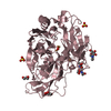
- Assembly
| Deposited unit | 
| ||||||||||||||||||
|---|---|---|---|---|---|---|---|---|---|---|---|---|---|---|---|---|---|---|---|
| 1 | 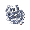
| ||||||||||||||||||
| 2 | 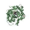
| ||||||||||||||||||
| Unit cell |
| ||||||||||||||||||
| Noncrystallographic symmetry (NCS) | NCS domain:
NCS domain segments: Component-ID: 1 / Ens-ID: 1 / Beg auth comp-ID: MET / Beg label comp-ID: MET / End auth comp-ID: HIS / End label comp-ID: HIS / Refine code: 5 / Auth seq-ID: 1 - 489 / Label seq-ID: 1 - 489
NCS oper: (Code: given Matrix: (0.44093, 0.8342, -0.33119), Vector : |
-Components
| #1: Protein | Mass: 56759.395 Da / Num. of mol.: 2 Source method: isolated from a genetically manipulated source Source: (gene. exp.) SULFOLOBUS SOLFATARICUS (archaea) / Production host:  ESCHERICHIA COLI (E. coli) / Strain (production host): BL21 / References: UniProt: P22498, beta-glucosidase ESCHERICHIA COLI (E. coli) / Strain (production host): BL21 / References: UniProt: P22498, beta-glucosidase#2: Chemical |   Mass: 305.349 Da / Num. of mol.: 2 / Source method: obtained synthetically / Formula: C16H21N2O4 Mass: 305.349 Da / Num. of mol.: 2 / Source method: obtained synthetically / Formula: C16H21N2O4#3: Chemical | Acetate  Mass: 59.044 Da / Num. of mol.: 3 / Source method: obtained synthetically / Formula: C2H3O2 Mass: 59.044 Da / Num. of mol.: 3 / Source method: obtained synthetically / Formula: C2H3O2#4: Water | ChemComp-HOH / | Water Mass: 18.015 Da / Num. of mol.: 741 / Source method: isolated from a natural source / Formula: H2O Mass: 18.015 Da / Num. of mol.: 741 / Source method: isolated from a natural source / Formula: H2O |
|---|
-Experimental details
-Experiment
| Experiment | Method: X-RAY DIFFRACTION / Number of used crystals: 1 |
|---|
- Sample preparation
| Crystal | Density Matthews: 3.4 Å3/Da / Density % sol: 63 % |
|---|---|
| Crystal grow | pH: 4.6 Details: 11-14% PEG4K, 0.1 M SODIUM ACETATE, 0.2 M AMMONIUM ACETATE, 10-13 MG/ML PROTEIN, 25% ETHYLENE GLYCOL, pH 4.60 |
-Data collection
| Diffraction | Mean temperature: 100 K |
|---|---|
| Diffraction source | Source: SYNCHROTRON / Site: ESRF  / Beamline: BM14 / Wavelength: 0.9793 / Beamline: BM14 / Wavelength: 0.9793 |
| Detector | Type: MARRESEARCH / Detector: CCD / Date: Jul 21, 2004 / Details: MIRRORS |
| Radiation | Monochromator: SI(111) / Protocol: SINGLE WAVELENGTH / Monochromatic (M) / Laue (L): M / Scattering type: x-ray |
| Radiation wavelength | Wavelength: 0.9793 Å / Relative weight: 1 |
| Reflection | Resolution: 2.3→30 Å / Num. obs: 68632 / % possible obs: 98.3 % / Redundancy: 6.1 % / Rmerge(I) obs: 0.11 / Net I/σ(I): 11.52 |
| Reflection shell | Resolution: 2.3→2.38 Å / Redundancy: 5.7 % / Rmerge(I) obs: 0.45 / Mean I/σ(I) obs: 3.48 / % possible all: 93.9 |
- Processing
| Software |
| ||||||||||||||||||||||||||||||||||||||||||||||||||||||||||||||||||||||||||||||||||||||||||||||||||||||||||||||||||||||||||||||||||||||||||||||||||||||||||||||||||||||||||||||||||||||
|---|---|---|---|---|---|---|---|---|---|---|---|---|---|---|---|---|---|---|---|---|---|---|---|---|---|---|---|---|---|---|---|---|---|---|---|---|---|---|---|---|---|---|---|---|---|---|---|---|---|---|---|---|---|---|---|---|---|---|---|---|---|---|---|---|---|---|---|---|---|---|---|---|---|---|---|---|---|---|---|---|---|---|---|---|---|---|---|---|---|---|---|---|---|---|---|---|---|---|---|---|---|---|---|---|---|---|---|---|---|---|---|---|---|---|---|---|---|---|---|---|---|---|---|---|---|---|---|---|---|---|---|---|---|---|---|---|---|---|---|---|---|---|---|---|---|---|---|---|---|---|---|---|---|---|---|---|---|---|---|---|---|---|---|---|---|---|---|---|---|---|---|---|---|---|---|---|---|---|---|---|---|---|---|
| Refinement | Method to determine structure: MOLECULAR REPLACEMENT Starting model: PDB ENTRY 1UWQ Resolution: 2.29→145.86 Å / Cor.coef. Fo:Fc: 0.958 / Cor.coef. Fo:Fc free: 0.935 / SU B: 6.712 / SU ML: 0.161 / Cross valid method: THROUGHOUT / ESU R: 0.264 / ESU R Free: 0.222 / Stereochemistry target values: MAXIMUM LIKELIHOOD / Details: HYDROGENS HAVE BEEN ADDED IN THE RIDING POSITIONS.
| ||||||||||||||||||||||||||||||||||||||||||||||||||||||||||||||||||||||||||||||||||||||||||||||||||||||||||||||||||||||||||||||||||||||||||||||||||||||||||||||||||||||||||||||||||||||
| Solvent computation | Ion probe radii: 0.8 Å / Shrinkage radii: 0.8 Å / VDW probe radii: 1.2 Å / Solvent model: MASK | ||||||||||||||||||||||||||||||||||||||||||||||||||||||||||||||||||||||||||||||||||||||||||||||||||||||||||||||||||||||||||||||||||||||||||||||||||||||||||||||||||||||||||||||||||||||
| Displacement parameters | Biso mean: 41.77 Å2
| ||||||||||||||||||||||||||||||||||||||||||||||||||||||||||||||||||||||||||||||||||||||||||||||||||||||||||||||||||||||||||||||||||||||||||||||||||||||||||||||||||||||||||||||||||||||
| Refinement step | Cycle: LAST / Resolution: 2.29→145.86 Å
| ||||||||||||||||||||||||||||||||||||||||||||||||||||||||||||||||||||||||||||||||||||||||||||||||||||||||||||||||||||||||||||||||||||||||||||||||||||||||||||||||||||||||||||||||||||||
| Refine LS restraints |
|