 Movie
Movie Controller

[English] 日本語
 Yorodumi
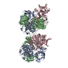
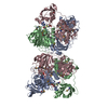
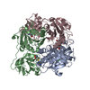
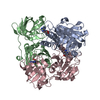
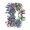
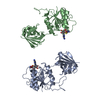
- PDB-2cc2: X-ray crystal structure of 5'-fluorodeoxyadenosine synthase from ... -
+ Open data

- Basic information
| Entry | Database: PDB / ID: 2cc2 | ||||||
|---|---|---|---|---|---|---|---|
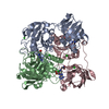
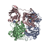
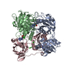
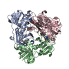
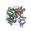
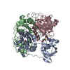
| Title | X-ray crystal structure of 5'-fluorodeoxyadenosine synthase from Streptomyces cattleya complexed with 5'deoxyadenosine | ||||||
 Components Components | 5'-FLUORO-5'-DEOXYADENOSINE SYNTHASE | ||||||
 Keywords Keywords |  TRANSFERASE / FLUORINASE / 5'DEOXYADENOSINE / FLA TRANSFERASE / FLUORINASE / 5'DEOXYADENOSINE / FLA | ||||||
| Function / homology |  Function and homology information Function and homology information | ||||||
| Biological species |  STREPTOMYCES CATTLEYA (bacteria) STREPTOMYCES CATTLEYA (bacteria) | ||||||
| Method | X-RAY DIFFRACTION / SYNCHROTRON / MOLECULAR REPLACEMENT / Resolution: 2 Å | ||||||
 Authors Authors | Mcewan, A.R. / Deng, H. / McGlinchey, R.P. / Robinson, D.R. / O'Hagan, D. / Spencer, J. / Naismith, J.H. | ||||||
 Citation Citation | Journal: Org. Biomol. Chem. / Year: 2006 Title: Substrate specificity in enzymatic fluorination. The fluorinase from Streptomyces cattleya accepts 2'-deoxyadenosine substrates. Authors: Cobb, S.L. / Deng, H. / McEwan, A.R. / Naismith, J.H. / O'Hagan, D. / Robinson, D.A. | ||||||
| History |
| ||||||
| Remark 700 | SHEET DETERMINATION METHOD: DSSP THE SHEETS PRESENTED AS "CB" IN EACH CHAIN ON SHEET RECORDS BELOW ... SHEET DETERMINATION METHOD: DSSP THE SHEETS PRESENTED AS "CB" IN EACH CHAIN ON SHEET RECORDS BELOW IS ACTUALLY AN 8-STRANDED BARREL THIS IS REPRESENTED BY A 9-STRANDED SHEET IN WHICH THE FIRST AND LAST STRANDS ARE IDENTICAL. THE SHEET STRUCTURE OF THIS MOLECULE IS BIFURCATED. IN ORDER TO REPRESENT THIS FEATURE IN THE SHEET RECORDS BELOW, TWO SHEETS ARE DEFINED. |
- Structure visualization
| Structure viewer | Molecule: MolmilJmol/JSmol |
|---|
- Downloads & links
-Download
| PDBx/mmCIF format | 2cc2.cif.gz | 333.8 KB | Display | PDBx/mmCIF format |
|---|---|---|---|---|
| PDB format | pdb2cc2.ent.gz | 270.6 KB | Display | PDB format |
| PDBx/mmJSON format | 2cc2.json.gz | Tree view | PDBx/mmJSON format | |
| Others |  Other downloads Other downloads |
-Validation report
| Arichive directory | https://data.pdbj.org/pub/pdb/validation_reports/cc/2cc2ftp://data.pdbj.org/pub/pdb/validation_reports/cc/2cc2 | HTTPS FTP |
|---|
-Related structure data
| Related structure data | 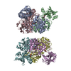 2c4uC  2c5bC 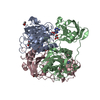 2cbxC  2c2wS S: Starting model for refinement C: citing same article ( |
|---|---|
| Similar structure data |


-Links
 PDBj
PDBj- Assembly
| Deposited unit | 
| ||||||||||||||||||||||||
|---|---|---|---|---|---|---|---|---|---|---|---|---|---|---|---|---|---|---|---|---|---|---|---|---|---|
| 1 | 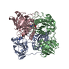
| ||||||||||||||||||||||||
| Unit cell |
| ||||||||||||||||||||||||
| Noncrystallographic symmetry (NCS) | NCS domain:
NCS domain segments: Component-ID: 1 / Ens-ID: 1 / Beg auth comp-ID: THR / Beg label comp-ID: THR / End auth comp-ID: GLU / End label comp-ID: GLU / Refine code: 6 / Auth seq-ID: 20 - 290 / Label seq-ID: 20 - 290
|
-Components
| #1: Protein | Mass: 32401.490 Da / Num. of mol.: 3 Source method: isolated from a genetically manipulated source Source: (gene. exp.) STREPTOMYCES CATTLEYA (bacteria) / Plasmid: PEHISTEV-FLA / Production host: Escherichia coli BL21(DE3) (bacteria) / References: UniProt: Q70GK9, adenosyl-fluoride synthase#2: Chemical | Deoxyadenosine  Mass: 251.242 Da / Num. of mol.: 3 / Source method: obtained synthetically / Formula: C10H13N5O3 Mass: 251.242 Da / Num. of mol.: 3 / Source method: obtained synthetically / Formula: C10H13N5O3#3: Chemical | ChemComp-CL / | Chloride  Mass: 35.453 Da / Num. of mol.: 1 / Source method: obtained synthetically / Formula: Cl Mass: 35.453 Da / Num. of mol.: 1 / Source method: obtained synthetically / Formula: Cl#4: Water | ChemComp-HOH / | Water Mass: 18.015 Da / Num. of mol.: 336 / Source method: isolated from a natural source / Formula: H2O Mass: 18.015 Da / Num. of mol.: 336 / Source method: isolated from a natural source / Formula: H2O |
|---|
-Experimental details
-Experiment
| Experiment | Method: X-RAY DIFFRACTION / Number of used crystals: 1 |
|---|
- Sample preparation
| Crystal | Density Matthews: 2.58 Å3/Da / Density % sol: 52.28 % |
|---|---|
| Crystal grow | pH: 4 Details: 24% PEG 1K, 100MM PHOSPHATE CITRATE PH4.4, 200MM LI2SO4, 20MM NACL, pH 4.00 |
-Data collection
| Diffraction | Mean temperature: 100 K |
|---|---|
| Diffraction source | Source: SYNCHROTRON / Site: ESRF  / Beamline: ID14-2 / Wavelength: 0.933 / Beamline: ID14-2 / Wavelength: 0.933 |
| Detector | Type: ADSC CCD / Detector: CCD / Date: May 21, 2005 / Details: TOROIDAL MIRROR |
| Radiation | Monochromator: DIAMOND 111-GE-20 / Protocol: SINGLE WAVELENGTH / Monochromatic (M) / Laue (L): M / Scattering type: x-ray |
| Radiation wavelength | Wavelength: 0.933 Å / Relative weight: 1 |
| Reflection | Resolution: 2→44.9 Å / Num. obs: 45862 / % possible obs: 74.1 % / Observed criterion σ(I): 2 / Redundancy: 2 % / Rmerge(I) obs: 0.08 / Net I/σ(I): 9.9 |
| Reflection shell | Resolution: 2→2.1 Å / Redundancy: 1.7 % / Rmerge(I) obs: 0.27 / Mean I/σ(I) obs: 2.1 / % possible all: 66.9 |
- Processing
| Software |
| ||||||||||||||||||||||||||||||||||||||||||||||||||||||||||||||||||||||||||||||||||||||||||||||||||||||||||||||||||||||||||||||||||||||||||||||||||||||||||||||||||||||||||||||||||||||
|---|---|---|---|---|---|---|---|---|---|---|---|---|---|---|---|---|---|---|---|---|---|---|---|---|---|---|---|---|---|---|---|---|---|---|---|---|---|---|---|---|---|---|---|---|---|---|---|---|---|---|---|---|---|---|---|---|---|---|---|---|---|---|---|---|---|---|---|---|---|---|---|---|---|---|---|---|---|---|---|---|---|---|---|---|---|---|---|---|---|---|---|---|---|---|---|---|---|---|---|---|---|---|---|---|---|---|---|---|---|---|---|---|---|---|---|---|---|---|---|---|---|---|---|---|---|---|---|---|---|---|---|---|---|---|---|---|---|---|---|---|---|---|---|---|---|---|---|---|---|---|---|---|---|---|---|---|---|---|---|---|---|---|---|---|---|---|---|---|---|---|---|---|---|---|---|---|---|---|---|---|---|---|---|
| Refinement | Method to determine structure: MOLECULAR REPLACEMENT Starting model: PDB ENTRY 2C2W Resolution: 2→35 Å / Cor.coef. Fo:Fc: 0.95 / Cor.coef. Fo:Fc free: 0.907 / SU B: 10.36 / SU ML: 0.131 / Cross valid method: THROUGHOUT / ESU R Free: 0.225 / Stereochemistry target values: MAXIMUM LIKELIHOOD Details: HYDROGENS HAVE BEEN ADDED IN THE RIDING POSITIONS. DISORDERED REGIONS WERE MODELED STEREOCHEMICALLY
| ||||||||||||||||||||||||||||||||||||||||||||||||||||||||||||||||||||||||||||||||||||||||||||||||||||||||||||||||||||||||||||||||||||||||||||||||||||||||||||||||||||||||||||||||||||||
| Solvent computation | Ion probe radii: 0.8 Å / Shrinkage radii: 0.8 Å / VDW probe radii: 1.4 Å / Solvent model: MASK | ||||||||||||||||||||||||||||||||||||||||||||||||||||||||||||||||||||||||||||||||||||||||||||||||||||||||||||||||||||||||||||||||||||||||||||||||||||||||||||||||||||||||||||||||||||||
| Displacement parameters | Biso mean: 26.08 Å2
| ||||||||||||||||||||||||||||||||||||||||||||||||||||||||||||||||||||||||||||||||||||||||||||||||||||||||||||||||||||||||||||||||||||||||||||||||||||||||||||||||||||||||||||||||||||||
| Refinement step | Cycle: LAST / Resolution: 2→35 Å
| ||||||||||||||||||||||||||||||||||||||||||||||||||||||||||||||||||||||||||||||||||||||||||||||||||||||||||||||||||||||||||||||||||||||||||||||||||||||||||||||||||||||||||||||||||||||
| Refine LS restraints |
|