 Movie
Movie Controller

[English] 日本語
 Yorodumi
- PDB-2c4m: Starch phosphorylase: structural studies explain oxyanion-depende... -
+ Open data

- Basic information
| Entry | Database: PDB / ID: 2c4m | ||||||
|---|---|---|---|---|---|---|---|
| Title | Starch phosphorylase: structural studies explain oxyanion-dependent kinetic stability and regulatory control. | ||||||
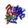
 Components Components | GLYCOGEN PHOSPHORYLASE | ||||||
 Keywords Keywords | TRANSFERASE / ALLOSTERIC CONTROL / PHOSPHATE DEPENDENCE / STARCH DEGRADING / PHOSPHORYLASE / GLYCOSYLTRANSFERASE | ||||||
| Function / homology |  Function and homology informationglycogen phosphorylase / glycogen phosphorylase activity / linear malto-oligosaccharide phosphorylase activity / SHG alpha-glucan phosphorylase activity / pyridoxal phosphate binding / carbohydrate metabolic process Function and homology informationglycogen phosphorylase / glycogen phosphorylase activity / linear malto-oligosaccharide phosphorylase activity / SHG alpha-glucan phosphorylase activity / pyridoxal phosphate binding / carbohydrate metabolic processSimilarity search - Function | ||||||
| Biological species |  CORYNEBACTERIUM CALLUNAE (bacteria) CORYNEBACTERIUM CALLUNAE (bacteria) | ||||||
| Method | X-RAY DIFFRACTION / SYNCHROTRON / MOLECULAR REPLACEMENT / Resolution: 1.9 Å | ||||||
 Authors Authors | Purvis, A. / Nidetzky, B. / Watson, K. | ||||||
 Citation Citation | Journal: To be Published Title: Starch Phosphorylase: Structural Studies Explain Oxyanion-Dependent Kinetic Stability and Regulatory Control Authors: Purvis, A. / Nidetzky, B. / Watson, K. | ||||||
| History |
| ||||||
| Remark 700 | SHEET THE SHEET STRUCTURE OF THIS MOLECULE IS BIFURCATED. IN ORDER TO REPRESENT THIS FEATURE IN ... SHEET THE SHEET STRUCTURE OF THIS MOLECULE IS BIFURCATED. IN ORDER TO REPRESENT THIS FEATURE IN THE SHEET RECORDS BELOW, TWO SHEETS ARE DEFINED. |
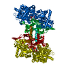
- Structure visualization
| Structure viewer | Molecule: MolmilJmol/JSmol |
|---|
- Downloads & links
-Download
| PDBx/mmCIF format | 2c4m.cif.gz | 645 KB | Display | PDBx/mmCIF format |
|---|---|---|---|---|
| PDB format | pdb2c4m.ent.gz | 528.9 KB | Display | PDB format |
| PDBx/mmJSON format | 2c4m.json.gz | Tree view | PDBx/mmJSON format | |
| Others |  Other downloads Other downloads |
-Validation report
| Arichive directory | https://data.pdbj.org/pub/pdb/validation_reports/c4/2c4mftp://data.pdbj.org/pub/pdb/validation_reports/c4/2c4m | HTTPS FTP |
|---|
-Related structure data
| Related structure data | 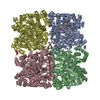 1gpaS S: Starting model for refinement |
|---|---|
| Similar structure data |


-Links
 PDBj
PDBj
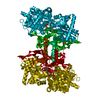
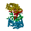
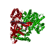
- Assembly
| Deposited unit | 
| ||||||||
|---|---|---|---|---|---|---|---|---|---|
| 1 | 
| ||||||||
| 2 | 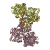
| ||||||||
| Unit cell |
|
-Components
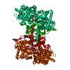
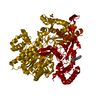
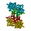
-Protein , 1 types, 4 molecules ABCD
| #1: Protein | / STARCH PHOSPHORYLASE Mass: 90662.922 Da / Num. of mol.: 4 / Mutation: YES Source method: isolated from a genetically manipulated source Details: PLP (1634) COFACTOR IS COVALENTLY LINKED TO LYS634 VIA SCHIFF BASE (C4A-NZ) Source: (gene. exp.) CORYNEBACTERIUM CALLUNAE (bacteria) / Plasmid: PQE 30 / Production host: Escherichia coli DH5[alpha] (bacteria) / Variant (production host): XL1 BLUE / References: UniProt: Q8KQ56, glycogen phosphorylase |
|---|
-Non-polymers , 5 types, 1189 molecules 


| #2: Chemical | ChemComp-PLP / Pyridoxal phosphate Mass: 247.142 Da / Num. of mol.: 4 / Source method: obtained synthetically / Formula: C8H10NO6P Mass: 247.142 Da / Num. of mol.: 4 / Source method: obtained synthetically / Formula: C8H10NO6P#3: Chemical | ChemComp-PO4 / Phosphate Mass: 94.971 Da / Num. of mol.: 9 / Source method: obtained synthetically / Formula: PO4 Mass: 94.971 Da / Num. of mol.: 9 / Source method: obtained synthetically / Formula: PO4#4: Chemical | ChemComp-FMT / Formic acid Mass: 46.025 Da / Num. of mol.: 19 / Source method: obtained synthetically / Formula: CH2O2 Mass: 46.025 Da / Num. of mol.: 19 / Source method: obtained synthetically / Formula: CH2O2#5: Chemical | Ethylene glycol Mass: 62.068 Da / Num. of mol.: 3 / Source method: obtained synthetically / Formula: C2H6O2 Mass: 62.068 Da / Num. of mol.: 3 / Source method: obtained synthetically / Formula: C2H6O2#6: Water | ChemComp-HOH / | WaterMass: 18.015 Da / Num. of mol.: 1154 / Source method: isolated from a natural source / Formula: H2O |
|---|
-Details
| Compound details | ENGINEERED RESIDUE IN CHAIN A, SER 225 TO ALA ENGINEERED RESIDUE IN CHAIN B, SER 225 TO ALA ...ENGINEERED |
|---|
-Experimental details
-Experiment
| Experiment | Method: X-RAY DIFFRACTION / Number of used crystals: 1 |
|---|
- Sample preparation
| Crystal | Density Matthews: 3.08 Å3/Da / Density % sol: 60 % Description: TWO SCANS WERE PERFORMED ON THE SAME CRYSTAL AT HIGH 1.9 ANG AND LOW 2.5 ANG RESOLUTION |
|---|---|
| Crystal grow | Method: vapor diffusion, hanging drop / pH: 5 Details: HANGING DROP VAPOUR DIFFUSION 8.4MG/ML PROTEIN SOLUTION, 0.1M SODIUM ACETATE PH 5.0, 8% PEG 8,000, 0.2M SODIUM FORMATE |
-Data collection
| Diffraction | Mean temperature: 100 K |
|---|---|
| Diffraction source | Source: SYNCHROTRON / Site: ESRF  / Beamline: ID14-1 / Wavelength: 0.934 / Beamline: ID14-1 / Wavelength: 0.934 |
| Detector | Type: ADSC CCD / Detector: CCD / Date: Mar 16, 2003 Details: SAGITALLY FOCUSING GE (220) CRYSTAL AND BENT MULTILAYER |
| Radiation | Monochromator: DIAMOND / Protocol: SINGLE WAVELENGTH / Monochromatic (M) / Laue (L): M / Scattering type: x-ray |
| Radiation wavelength | Wavelength: 0.934 Å / Relative weight: 1 |
| Reflection | Resolution: 1.9→30 Å / Num. obs: 306185 / % possible obs: 89.7 % / Observed criterion σ(I): 0 / Redundancy: 2.6 % / Biso Wilson estimate: 18.5 Å2 / Rmerge(I) obs: 0.08 / Net I/σ(I): 5.1 |
| Reflection shell | Resolution: 1.9→2 Å / Redundancy: 2 % / Rmerge(I) obs: 0.24 / Mean I/σ(I) obs: 3.1 / % possible all: 75.9 |
- Processing
| Software |
| ||||||||||||||||||||||||||||||||||||||||||||||||||||||||||||||||||||||||||||||||
|---|---|---|---|---|---|---|---|---|---|---|---|---|---|---|---|---|---|---|---|---|---|---|---|---|---|---|---|---|---|---|---|---|---|---|---|---|---|---|---|---|---|---|---|---|---|---|---|---|---|---|---|---|---|---|---|---|---|---|---|---|---|---|---|---|---|---|---|---|---|---|---|---|---|---|---|---|---|---|---|---|---|
| Refinement | Method to determine structure: MOLECULAR REPLACEMENT Starting model: PDB ENTRY 1GPA Resolution: 1.9→30 Å / Rfactor Rfree error: 0.002 / Data cutoff high absF: 3314122.3 / Isotropic thermal model: RESTRAINED / σ(F): 0 Details: INITIAL REFINEMENT WAS CARRIED OUT USING 4 FOLD NCS RESTRAINTS. IN THE REFINEMENT THE FOLLOWING RESIDUES WERE GIVEN ZERO OCCUPANCY DUE TO THE ABSENCE OF ELECTRON DENSITY. CHAIN ID A Q5, R43, ...Details: INITIAL REFINEMENT WAS CARRIED OUT USING 4 FOLD NCS RESTRAINTS. IN THE REFINEMENT THE FOLLOWING RESIDUES WERE GIVEN ZERO OCCUPANCY DUE TO THE ABSENCE OF ELECTRON DENSITY. CHAIN ID A Q5, R43, K281, E319, K327, V336, L337, E339, E378, E505, E510, D647, E659, K724, R775, R778. CHAIN ID B K281, E319, E339, E378, E501, E505, E510, E659, E677, N707, K724, E741, R775, R778. CHAIN ID C Q5, R92, E151, K281, D296, E339, E378, K474, K482, E505, R523, E546, D551, K724, K794. CHAIN ID D Q5, R43, E86, R92, E97, E151, K281, E319, E339, E378, E505, E677, K680, K724, R775, R778.
| ||||||||||||||||||||||||||||||||||||||||||||||||||||||||||||||||||||||||||||||||
| Solvent computation | Solvent model: FLAT MODEL / Bsol: 38.6 Å2 / ksol: 0.34 e/Å3 | ||||||||||||||||||||||||||||||||||||||||||||||||||||||||||||||||||||||||||||||||
| Displacement parameters | Biso mean: 31.2 Å2
| ||||||||||||||||||||||||||||||||||||||||||||||||||||||||||||||||||||||||||||||||
| Refine analyze |
| ||||||||||||||||||||||||||||||||||||||||||||||||||||||||||||||||||||||||||||||||
| Refinement step | Cycle: LAST / Resolution: 1.9→30 Å
| ||||||||||||||||||||||||||||||||||||||||||||||||||||||||||||||||||||||||||||||||
| Refine LS restraints |
| ||||||||||||||||||||||||||||||||||||||||||||||||||||||||||||||||||||||||||||||||
| LS refinement shell | Resolution: 1.9→2.02 Å / Rfactor Rfree error: 0.007 / Total num. of bins used: 6
| ||||||||||||||||||||||||||||||||||||||||||||||||||||||||||||||||||||||||||||||||
| Xplor file |
|
