 Movie
Movie Controller

[English] 日本語
 Yorodumi
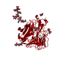
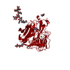
- PDB-2c4a: Structure of Neuraminidase Subtype N9 Complexed with 30 MM Sialic... -
+ Open data

- Basic information
| Entry | Database: PDB / ID: 2c4a | |||||||||
|---|---|---|---|---|---|---|---|---|---|---|
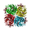
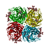
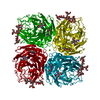
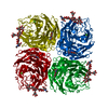
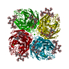
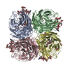
| Title | Structure of Neuraminidase Subtype N9 Complexed with 30 MM Sialic Acid (NANA, NEU5AC), Crystal Soaked for 3 Hours at 291 K. | |||||||||
 Components Components | NEURAMINIDASE SUBTYPE N9 | |||||||||
 Keywords Keywords |  HYDROLASE / INFLUENZA TYPE A / NEURAMINIDASE / SIALIC ACID / SUBTYPE N9 / GLYCOPROTEIN / GLYCOSIDASE / TRANSMEMBRANE HYDROLASE / INFLUENZA TYPE A / NEURAMINIDASE / SIALIC ACID / SUBTYPE N9 / GLYCOPROTEIN / GLYCOSIDASE / TRANSMEMBRANE | |||||||||
| Function / homology |  Function and homology information Function and homology informationexo-alpha-(2->3)-sialidase activity / exo-alpha-(2->6)-sialidase activity / exo-alpha-(2->8)-sialidase activity / exo-alpha-sialidase / viral budding from plasma membrane / carbohydrate metabolic process / host cell plasma membrane / virion membrane / membrane / metal ion bindingSimilarity search - Function | |||||||||
| Biological species |   INFLUENZA A VIRUS INFLUENZA A VIRUS | |||||||||
| Method | X-RAY DIFFRACTION / MOLECULAR REPLACEMENT / Resolution: 2.15 Å | |||||||||
 Authors Authors | Rudino-Pinera, E. / Tunnah, P. / Crennell, S.J. / Webster, R.G. / Laver, W.G. / Garman, E.F. | |||||||||
 Citation Citation | Journal: To be Published Title: The Crystal Structure of Influenza Type a Virus Neuraminidase of the N6 Subtype at 1.85 A Resolution Authors: Rudino-Pinera, E. / Crennell, S.J. / Webster, R.G. / Laver, W.G. / Garman, E.F. | |||||||||
| History |
|
- Structure visualization
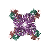
| Structure viewer | Molecule: MolmilJmol/JSmol |
|---|
- Downloads & links
-Download
| PDBx/mmCIF format | 2c4a.cif.gz | 104.8 KB | Display | PDBx/mmCIF format |
|---|---|---|---|---|
| PDB format | pdb2c4a.ent.gz | 78.6 KB | Display | PDB format |
| PDBx/mmJSON format | 2c4a.json.gz | Tree view | PDBx/mmJSON format | |
| Others |  Other downloads Other downloads |
-Validation report
| Arichive directory | https://data.pdbj.org/pub/pdb/validation_reports/c4/2c4aftp://data.pdbj.org/pub/pdb/validation_reports/c4/2c4a | HTTPS FTP |
|---|
-Related structure data
| Related structure data | 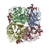 1v0zC  2c4lC  7nn9S S: Starting model for refinement C: citing same article ( |
|---|---|
| Similar structure data |


-Links
 PDBj
PDBj

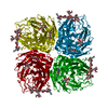
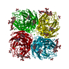
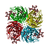
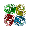
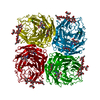
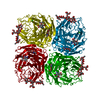
- Assembly
| Deposited unit | 
| ||||||||
|---|---|---|---|---|---|---|---|---|---|
| 1 | 
| ||||||||
| Unit cell |
| ||||||||
| Components on special symmetry positions |
|
-Components
-Protein , 1 types, 1 molecules A
| #1: Protein | Mass: 43723.770 Da / Num. of mol.: 1 / Fragment: RESIDUES 83-470 Source method: isolated from a genetically manipulated source Source: (gene. exp.) INFLUENZA A VIRUS / Strain: A/TERN/STERNA ALBIFRONS/N9 / Production host:  GALLUS GALLUS (chicken) / References: UniProt: P03472, exo-alpha-sialidase GALLUS GALLUS (chicken) / References: UniProt: P03472, exo-alpha-sialidase |
|---|
-Sugars , 4 types, 5 molecules 

| #2: Polysaccharide | alpha-D-mannopyranose-(1-2)-alpha-D-mannopyranose-(1-2)-alpha-D-mannopyranose-(1-3)-[alpha-D- ...alpha-D-mannopyranose-(1-2)-alpha-D-mannopyranose-(1-2)-alpha-D-mannopyranose-(1-3)-[alpha-D-mannopyranose-(1-3)-[alpha-D-mannopyranose-(1-6)]alpha-D-mannopyranose-(1-6)]beta-D-mannopyranose-(1-4)-2-acetamido-2-deoxy-beta-D-glucopyranose-(1-4)-2-acetamido-2-deoxy-beta-D-glucopyranose / Mass: 1559.386 Da / Num. of mol.: 1 Source method: isolated from a genetically manipulated source | ||
|---|---|---|---|
| #3: Polysaccharide | 2-acetamido-2-deoxy-beta-D-glucopyranose-(1-4)-2-acetamido-2-deoxy-beta-D-glucopyranose / Mass: 424.401 Da / Num. of mol.: 1 Source method: isolated from a genetically manipulated source | ||
| #4: Sugar | Sialic acid Type: D-saccharide, alpha linking / Mass: 309.270 Da / Num. of mol.: 2 Type: D-saccharide, alpha linking / Mass: 309.270 Da / Num. of mol.: 2Source method: isolated from a genetically manipulated source Formula: C11H19NO9 #5: Sugar | ChemComp-NAG / | N-Acetylglucosamine Type: D-saccharide, beta linking / Mass: 221.208 Da / Num. of mol.: 1 Type: D-saccharide, beta linking / Mass: 221.208 Da / Num. of mol.: 1Source method: isolated from a genetically manipulated source Formula: C8H15NO6 |
-Non-polymers , 2 types, 260 molecules 

| #6: Chemical | ChemComp-CA /  Mass: 40.078 Da / Num. of mol.: 1 / Source method: obtained synthetically / Formula: Ca Mass: 40.078 Da / Num. of mol.: 1 / Source method: obtained synthetically / Formula: Ca |
|---|---|
| #7: Water | ChemComp-HOH / WaterMass: 18.015 Da / Num. of mol.: 259 / Source method: isolated from a natural source / Formula: H2O |
-Experimental details
-Experiment
| Experiment | Method: X-RAY DIFFRACTION / Number of used crystals: 1 |
|---|
- Sample preparation
| Crystal | Density Matthews: 2.94 Å3/Da / Density % sol: 58.22 % |
|---|---|
| Crystal grow | pH: 6.8 Details: PROTEIN WAS CRYSTALLISED FROM 1.9M POTASSIUM PHOSPHATE PH 6.8, THEN SOAKED FOR 3 HOURS IN 30 MM NEU5AC |
-Data collection
| Diffraction | Mean temperature: 100 K |
|---|---|
| Diffraction source | Source: ROTATING ANODE / Type: RIGAKU RU200H / Wavelength: 1.5418 |
| Detector | Type: MAR scanner 345 mm plate / Detector: IMAGE PLATE / Date: Jun 10, 2004 / Details: CONFOCAL OSMIC BLUE |
| Radiation | Protocol: SINGLE WAVELENGTH / Monochromatic (M) / Laue (L): M / Scattering type: x-ray |
| Radiation wavelength | Wavelength: 1.5418 Å / Relative weight: 1 |
| Reflection | Resolution: 2.15→38.66 Å / Num. obs: 27874 / % possible obs: 100 % / Observed criterion σ(I): 0 / Redundancy: 9.9 % / Biso Wilson estimate: 16.57 Å2 / Rmerge(I) obs: 0.12 / Net I/σ(I): 5.5 |
| Reflection shell | Resolution: 2.15→2.21 Å / Redundancy: 9.4 % / Rmerge(I) obs: 0.27 / Mean I/σ(I) obs: 2.8 / % possible all: 100 |
- Processing
| Software |
| ||||||||||||||||||||||||||||||||||||||||||||||||||||||||||||||||||||||||||||||||||||||||||||||||||||||||||||||||||||||||||||||||||||||||||||||||||||||||||||||||||||||||||||||||||||||
|---|---|---|---|---|---|---|---|---|---|---|---|---|---|---|---|---|---|---|---|---|---|---|---|---|---|---|---|---|---|---|---|---|---|---|---|---|---|---|---|---|---|---|---|---|---|---|---|---|---|---|---|---|---|---|---|---|---|---|---|---|---|---|---|---|---|---|---|---|---|---|---|---|---|---|---|---|---|---|---|---|---|---|---|---|---|---|---|---|---|---|---|---|---|---|---|---|---|---|---|---|---|---|---|---|---|---|---|---|---|---|---|---|---|---|---|---|---|---|---|---|---|---|---|---|---|---|---|---|---|---|---|---|---|---|---|---|---|---|---|---|---|---|---|---|---|---|---|---|---|---|---|---|---|---|---|---|---|---|---|---|---|---|---|---|---|---|---|---|---|---|---|---|---|---|---|---|---|---|---|---|---|---|---|
| Refinement | Method to determine structure: MOLECULAR REPLACEMENT Starting model: PDB ENTRY 7NN9 Resolution: 2.15→30.1 Å / Cor.coef. Fo:Fc: 0.95 / Cor.coef. Fo:Fc free: 0.924 / SU B: 3.66 / SU ML: 0.098 / Cross valid method: THROUGHOUT / ESU R: 0.184 / ESU R Free: 0.164 / Stereochemistry target values: MAXIMUM LIKELIHOOD
| ||||||||||||||||||||||||||||||||||||||||||||||||||||||||||||||||||||||||||||||||||||||||||||||||||||||||||||||||||||||||||||||||||||||||||||||||||||||||||||||||||||||||||||||||||||||
| Solvent computation | Ion probe radii: 0.8 Å / Shrinkage radii: 0.8 Å / VDW probe radii: 1.2 Å / Solvent model: MASK | ||||||||||||||||||||||||||||||||||||||||||||||||||||||||||||||||||||||||||||||||||||||||||||||||||||||||||||||||||||||||||||||||||||||||||||||||||||||||||||||||||||||||||||||||||||||
| Displacement parameters | Biso mean: 12.2 Å2 | ||||||||||||||||||||||||||||||||||||||||||||||||||||||||||||||||||||||||||||||||||||||||||||||||||||||||||||||||||||||||||||||||||||||||||||||||||||||||||||||||||||||||||||||||||||||
| Refinement step | Cycle: LAST / Resolution: 2.15→30.1 Å
| ||||||||||||||||||||||||||||||||||||||||||||||||||||||||||||||||||||||||||||||||||||||||||||||||||||||||||||||||||||||||||||||||||||||||||||||||||||||||||||||||||||||||||||||||||||||
| Refine LS restraints |
|