 Movie
Movie Controller

[English] 日本語
 Yorodumi
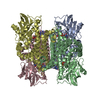
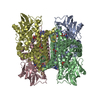
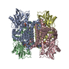
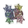
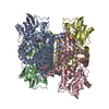
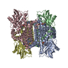
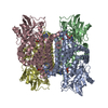
- PDB-2c12: Crystal Structure of Nitroalkane Oxidase in Complex with Spermine... -
+ Open data

- Basic information
| Entry | Database: PDB / ID: 2c12 | ||||||
|---|---|---|---|---|---|---|---|
| Title | Crystal Structure of Nitroalkane Oxidase in Complex with Spermine, a Competitive Inhibitor | ||||||
 Components Components | NITROALKANE OXIDASE | ||||||
 Keywords Keywords | OXIDOREDUCTASE / FLAVOENZYME / NITROALKANE / ACYL-COA DEHYDROGENASE / LONG CELL EDGE / FAD / INHIBITOR / FLAVOPROTEIN | ||||||
| Function / homology |  Function and homology informationnitroalkane oxidase activity / nitroalkane oxidase / nitroethane oxidase activity / butyrate catabolic process / fatty acid beta-oxidation using acyl-CoA dehydrogenase / oxidoreductase activity, acting on the CH-CH group of donors / : / FAD binding Function and homology informationnitroalkane oxidase activity / nitroalkane oxidase / nitroethane oxidase activity / butyrate catabolic process / fatty acid beta-oxidation using acyl-CoA dehydrogenase / oxidoreductase activity, acting on the CH-CH group of donors / : / FAD bindingSimilarity search - Function | ||||||
| Biological species |   FUSARIUM OXYSPORUM (fungus) FUSARIUM OXYSPORUM (fungus) | ||||||
| Method | X-RAY DIFFRACTION / SYNCHROTRON / MOLECULAR REPLACEMENT / Resolution: 2.07 Å | ||||||
 Authors Authors | Nagpal, A. / Valley, M.P. / Fitzpatrick, P.F. / Orville, A.M. | ||||||
 Citation Citation | Journal: Biochemistry / Year: 2006 Title: Crystal Structures of Nitroalkane Oxidase: Insights Into the Reaction Mechanism from a Covalent Complex of the Flavoenzyme Trapped During Turnover. Authors: Nagpal, A. / Valley, M.P. / Fitzpatrick, P.F. / Orville, A.M. #1: Journal: Acta Crystallogr.,Sect.D / Year: 2004 Title: Crystallization and Preliminary Analysis of Active Nitroalkane Oxidase in Three Crystal Forms Authors: Nagpal, A. / Valley, M.P. / Fitzpatrick, P.F. / Orville, A.M. #2: Journal: Arch.Biochem.Biophys. / Year: 2005 Title: Nitroalkane Oxidase, a Carbanion-Forming Flavoprotein Homologous to Acyl-Coa Dehydrogenase Authors: Fitzpatrick, P.F. / Orville, A.M. / Nagpal, A. / Valley, M.P. | ||||||
| History |
| ||||||
| Remark 650 | HELIX DETERMINATION METHOD: AUTHOR PROVIDED. | ||||||
| Remark 700 | SHEET DETERMINATION METHOD: AUTHOR PROVIDED. |
- Structure visualization
| Structure viewer | Molecule: MolmilJmol/JSmol |
|---|
- Downloads & links
-Download
| PDBx/mmCIF format | 2c12.cif.gz | 522.8 KB | Display | PDBx/mmCIF format |
|---|---|---|---|---|
| PDB format | pdb2c12.ent.gz | 430.7 KB | Display | PDB format |
| PDBx/mmJSON format | 2c12.json.gz | Tree view | PDBx/mmJSON format | |
| Others |  Other downloads Other downloads |
-Validation report
| Arichive directory | https://data.pdbj.org/pub/pdb/validation_reports/c1/2c12ftp://data.pdbj.org/pub/pdb/validation_reports/c1/2c12 | HTTPS FTP |
|---|
-Related structure data
| Related structure data |  2c0uSC S: Starting model for refinement C: citing same article ( |
|---|---|
| Similar structure data |
-Links
 PDBj
PDBj

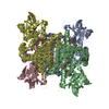
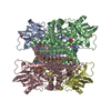
- Assembly
| Deposited unit | 
| ||||||||
|---|---|---|---|---|---|---|---|---|---|
| 1 | 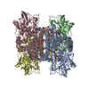
| ||||||||
| 2 | 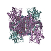
| ||||||||
| Unit cell |
|
-Components
-Protein , 1 types, 6 molecules ABCDEF
| #1: Protein | Mass: 48219.246 Da / Num. of mol.: 6 Source method: isolated from a genetically manipulated source Source: (gene. exp.) FUSARIUM OXYSPORUM (fungus) / Plasmid: PETNAO4 / Production host:  ESCHERICHIA COLI (E. coli) / Strain (production host): BL21(DE3) / References: UniProt: Q8X1D8 ESCHERICHIA COLI (E. coli) / Strain (production host): BL21(DE3) / References: UniProt: Q8X1D8 |
|---|
-Non-polymers , 5 types, 1144 molecules 


| #2: Chemical | ChemComp-FAD / Flavin adenine dinucleotide Mass: 785.550 Da / Num. of mol.: 6 / Source method: obtained synthetically / Formula: C27H33N9O15P2 / Comment: FAD*YM Mass: 785.550 Da / Num. of mol.: 6 / Source method: obtained synthetically / Formula: C27H33N9O15P2 / Comment: FAD*YM#3: Chemical | ChemComp-GOL / Glycerol Mass: 92.094 Da / Num. of mol.: 15 / Source method: obtained synthetically / Formula: C3H8O3 Mass: 92.094 Da / Num. of mol.: 15 / Source method: obtained synthetically / Formula: C3H8O3#4: Chemical | ChemComp-SPM / Spermine Mass: 202.340 Da / Num. of mol.: 4 / Source method: obtained synthetically / Formula: C10H26N4 Mass: 202.340 Da / Num. of mol.: 4 / Source method: obtained synthetically / Formula: C10H26N4#5: Chemical | Polyethylene glycol Mass: 354.436 Da / Num. of mol.: 2 / Source method: obtained synthetically / Formula: C16H34O8 / Comment: precipitant*YM Mass: 354.436 Da / Num. of mol.: 2 / Source method: obtained synthetically / Formula: C16H34O8 / Comment: precipitant*YM#6: Water | ChemComp-HOH / | WaterMass: 18.015 Da / Num. of mol.: 1117 / Source method: isolated from a natural source / Formula: H2O |
|---|
-Experimental details
-Experiment
| Experiment | Method: X-RAY DIFFRACTION / Number of used crystals: 3 |
|---|
- Sample preparation
| Crystal | Density Matthews: 2.58 Å3/Da / Density % sol: 52.2 % |
|---|---|
| Crystal grow | pH: 7.5 Details: 25%(W/V) PEG 4000, 35% (V/V) GLYCEROL, 200 MM SODIUM CACODYLATE TRIHYDRATE PH 7.5, 1 MM SPERMINE TETRAHYDROCHLORIDE |
-Data collection
| Diffraction | Mean temperature: 100 K |
|---|---|
| Diffraction source | Source: SYNCHROTRON / Site: APS  / Beamline: 14-BM-C / Wavelength: 0.9 / Beamline: 14-BM-C / Wavelength: 0.9 |
| Detector | Type: ADSC CCD / Detector: CCD / Date: Aug 9, 2003 |
| Radiation | Protocol: SINGLE WAVELENGTH / Monochromatic (M) / Laue (L): M / Scattering type: x-ray |
| Radiation wavelength | Wavelength: 0.9 Å / Relative weight: 1 |
| Reflection | Resolution: 2.07→50 Å / Num. obs: 180533 / % possible obs: 97.8 % / Observed criterion σ(I): 3 / Redundancy: 1.64 % / Rmerge(I) obs: 0.14 / Net I/σ(I): 12.6 |
| Reflection shell | Resolution: 2.07→2.15 Å / Redundancy: 1 % / Rmerge(I) obs: 0.269 / Mean I/σ(I) obs: 3 / % possible all: 81.9 |
- Processing
| Software |
| ||||||||||||||||||||||||||||||||||||||||||||||||||||||||||||||||||||||||||||||||||||||||||||||||||||||||||||||||||||||||||||||||||||||||||||||||||||||||||||||||||||||||||||||||||||||
|---|---|---|---|---|---|---|---|---|---|---|---|---|---|---|---|---|---|---|---|---|---|---|---|---|---|---|---|---|---|---|---|---|---|---|---|---|---|---|---|---|---|---|---|---|---|---|---|---|---|---|---|---|---|---|---|---|---|---|---|---|---|---|---|---|---|---|---|---|---|---|---|---|---|---|---|---|---|---|---|---|---|---|---|---|---|---|---|---|---|---|---|---|---|---|---|---|---|---|---|---|---|---|---|---|---|---|---|---|---|---|---|---|---|---|---|---|---|---|---|---|---|---|---|---|---|---|---|---|---|---|---|---|---|---|---|---|---|---|---|---|---|---|---|---|---|---|---|---|---|---|---|---|---|---|---|---|---|---|---|---|---|---|---|---|---|---|---|---|---|---|---|---|---|---|---|---|---|---|---|---|---|---|---|
| Refinement | Method to determine structure: MOLECULAR REPLACEMENT Starting model: PDB ENTRY 2C0U Resolution: 2.07→50 Å / Cor.coef. Fo:Fc: 0.939 / Cor.coef. Fo:Fc free: 0.915 / SU B: 4.182 / SU ML: 0.112 / Cross valid method: THROUGHOUT / ESU R: 0.2 / ESU R Free: 0.169 / Stereochemistry target values: MAXIMUM LIKELIHOOD / Details: HYDROGENS HAVE BEEN ADDED IN THE RIDING POSITIONS.
| ||||||||||||||||||||||||||||||||||||||||||||||||||||||||||||||||||||||||||||||||||||||||||||||||||||||||||||||||||||||||||||||||||||||||||||||||||||||||||||||||||||||||||||||||||||||
| Solvent computation | Ion probe radii: 0.8 Å / Shrinkage radii: 0.8 Å / VDW probe radii: 1.4 Å / Solvent model: BABINET MODEL WITH MASK | ||||||||||||||||||||||||||||||||||||||||||||||||||||||||||||||||||||||||||||||||||||||||||||||||||||||||||||||||||||||||||||||||||||||||||||||||||||||||||||||||||||||||||||||||||||||
| Displacement parameters | Biso mean: 24.66 Å2
| ||||||||||||||||||||||||||||||||||||||||||||||||||||||||||||||||||||||||||||||||||||||||||||||||||||||||||||||||||||||||||||||||||||||||||||||||||||||||||||||||||||||||||||||||||||||
| Refinement step | Cycle: LAST / Resolution: 2.07→50 Å
| ||||||||||||||||||||||||||||||||||||||||||||||||||||||||||||||||||||||||||||||||||||||||||||||||||||||||||||||||||||||||||||||||||||||||||||||||||||||||||||||||||||||||||||||||||||||
| Refine LS restraints |
|