 Movie
Movie Controller


+ Open data

- Basic information
| Entry | Database: PDB / ID: 2bzx | ||||||
|---|---|---|---|---|---|---|---|
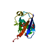
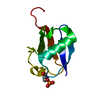
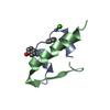
| Title | Atomic model of CrkL-SH3C monomer | ||||||
 Components Components | CRK-LIKE PROTEIN | ||||||
 Keywords Keywords |  SH3 DOMAIN / CRKL / SH3C / MONOMER / NATIVE / NUCLEAR EXPORT SH3 DOMAIN / CRKL / SH3C / MONOMER / NATIVE / NUCLEAR EXPORT | ||||||
| Function / homology |  Function and homology information Function and homology informationpositive regulation of glial cell migration / chordate pharynx development / extrinsic component of postsynaptic membrane / helper T cell diapedesis / cerebellar neuron development / postsynaptic specialization assembly / urogenital system development / parathyroid gland development / regulation of T cell migration / reelin-mediated signaling pathway ...positive regulation of glial cell migration / chordate pharynx development / extrinsic component of postsynaptic membrane / helper T cell diapedesis / cerebellar neuron development / postsynaptic specialization assembly / urogenital system development / parathyroid gland development / regulation of T cell migration / reelin-mediated signaling pathway / endothelin receptor signaling pathway / regulation of dendrite development / positive regulation of skeletal muscle acetylcholine-gated channel clustering / cranial skeletal system development / B cell apoptotic process / MET receptor recycling / cellular response to interleukin-7 / acetylcholine receptor signaling pathway / MET activates RAP1 and RAC1 / anterior/posterior pattern specification / Frs2-mediated activation / establishment of cell polarity / blood vessel development / dendrite development / outflow tract morphogenesis / positive regulation of Rac protein signal transduction / regulation of cell adhesion mediated by integrin / single fertilization / retinoic acid receptor signaling pathway / fibroblast growth factor receptor signaling pathway / signaling adaptor activity / Erythropoietin activates RAS / positive regulation of substrate adhesion-dependent cell spreading / JNK cascade / cellular response to transforming growth factor beta stimulus / phosphotyrosine residue binding / cell chemotaxis / Downstream signal transduction / thymus development / negative regulation of protein phosphorylation / Regulation of signaling by CBL / regulation of cell growth / hippocampus development / neuron migration / neuromuscular junction / lipid metabolic process / receptor tyrosine kinase binding / cerebral cortex development / male gonad development / cell migration / cellular response to xenobiotic stimulus / T cell receptor signaling pathway / spermatogenesis / RNA polymerase II-specific DNA-binding transcription factor binding / Ras protein signal transduction / positive regulation of ERK1 and ERK2 cascade / intracellular signal transduction / cadherin binding / positive regulation of protein phosphorylation / negative regulation of gene expression / positive regulation of cell population proliferation / signal transduction / protein-containing complex / RNA binding / nucleoplasm / identical protein binding / cytosolSimilarity search - Function | ||||||
| Biological species |  HOMO SAPIENS (human) HOMO SAPIENS (human) | ||||||
| Method | X-RAY DIFFRACTION / SYNCHROTRON / MOLECULAR REPLACEMENT / Resolution: 2.8 Å | ||||||
 Authors Authors | Harkiolaki, M. / Gilbert, R.J. / Jones, E.Y. / Feller, S.M. | ||||||
 Citation Citation | Journal: Structure / Year: 2006 Title: The C-Terminal SH3 Domain of Crkl as a Dynamic Dimerization Module Transiently Exposing a Nuclear Export Signal. Authors: Harkiolaki, M. / Gilbert, R.J. / Jones, E.Y. / Feller, S.M. | ||||||
| History |
|
- Structure visualization
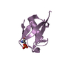
| Structure viewer | Molecule: MolmilJmol/JSmol |
|---|
- Downloads & links
-Download
| PDBx/mmCIF format | 2bzx.cif.gz | 23.9 KB | Display | PDBx/mmCIF format |
|---|---|---|---|---|
| PDB format | pdb2bzx.ent.gz | 14.6 KB | Display | PDB format |
| PDBx/mmJSON format | 2bzx.json.gz | Tree view | PDBx/mmJSON format | |
| Others |  Other downloads Other downloads |
-Validation report
| Arichive directory | https://data.pdbj.org/pub/pdb/validation_reports/bz/2bzxftp://data.pdbj.org/pub/pdb/validation_reports/bz/2bzx | HTTPS FTP |
|---|
-Related structure data
| Related structure data | 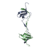 2bzyC 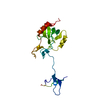 1uecS C: citing same article ( S: Starting model for refinement |
|---|---|
| Similar structure data |


-Links
 PDBj
PDBj


- Assembly
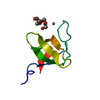
| Deposited unit | 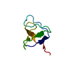
| ||||||||
|---|---|---|---|---|---|---|---|---|---|
| 1 |
| ||||||||
| Unit cell |
|
-Components
| #1: Protein | Mass: 7551.613 Da / Num. of mol.: 1 / Fragment: SH3 DOMAIN Source method: isolated from a genetically manipulated source Source: (gene. exp.) HOMO SAPIENS (human) / Plasmid: PGEX-KG / Production host:  ESCHERICHIA COLI (E. coli) / Strain (production host): TOPP1 / References: UniProt: P46109 ESCHERICHIA COLI (E. coli) / Strain (production host): TOPP1 / References: UniProt: P46109 |
|---|---|
| Compound details | MAY MEDIATE THE TRANSDUCTI |
-Experimental details
-Experiment
| Experiment | Method: X-RAY DIFFRACTION / Number of used crystals: 1 |
|---|
- Sample preparation
| Crystal | Density Matthews: 2.75 Å3/Da / Density % sol: 54.91 % |
|---|---|
| Crystal grow | pH: 6.5 Details: 1.7M AMMONIUM SULPHATE, 25.5% W/V PEG 8000, 0.085M SODIUM CACODYLATE PH6.5, 15% V/V GLYCEROL, pH 6.50 |
-Data collection
| Diffraction | Mean temperature: 100 K |
|---|---|
| Diffraction source | Source: SYNCHROTRON / Site: ESRF  / Beamline: BM14 / Wavelength: 1.7712 / Beamline: BM14 / Wavelength: 1.7712 |
| Detector | Type: MARRESEARCH / Detector: CCD / Date: Oct 8, 2003 / Details: MIRRORS |
| Radiation | Monochromator: SI 111 / Protocol: SINGLE WAVELENGTH / Monochromatic (M) / Laue (L): M / Scattering type: x-ray |
| Radiation wavelength | Wavelength: 1.7712 Å / Relative weight: 1 |
| Reflection | Resolution: 2.8→30 Å / Num. obs: 2035 / % possible obs: 99.6 % / Observed criterion σ(I): 0 / Redundancy: 38.8 % / Biso Wilson estimate: 87 Å2 / Rmerge(I) obs: 0.1 / Net I/σ(I): 34.3 |
| Reflection shell | Resolution: 2.8→2.9 Å / Redundancy: 26.8 % / Rmerge(I) obs: 1 / Mean I/σ(I) obs: 3.2 / % possible all: 99.5 |
- Processing
| Software |
| ||||||||||||||||||||||||||||||||||||||||||||||||||||||||||||||||||||||||||||||||
|---|---|---|---|---|---|---|---|---|---|---|---|---|---|---|---|---|---|---|---|---|---|---|---|---|---|---|---|---|---|---|---|---|---|---|---|---|---|---|---|---|---|---|---|---|---|---|---|---|---|---|---|---|---|---|---|---|---|---|---|---|---|---|---|---|---|---|---|---|---|---|---|---|---|---|---|---|---|---|---|---|---|
| Refinement | Method to determine structure: MOLECULAR REPLACEMENT Starting model: PDB ENTRY 1UEC Resolution: 2.8→20 Å / Data cutoff high absF: 10000 / Isotropic thermal model: RESTRAINED / Cross valid method: FREE R-VALUE / σ(F): 1 / Stereochemistry target values: MAXIMUM LIKELIHOOD
| ||||||||||||||||||||||||||||||||||||||||||||||||||||||||||||||||||||||||||||||||
| Solvent computation | Solvent model: CNS BULK SOLVENT CORRECTION / Bsol: 60 Å2 / ksol: 0.32 e/Å3 | ||||||||||||||||||||||||||||||||||||||||||||||||||||||||||||||||||||||||||||||||
| Displacement parameters | Biso mean: 73.3 Å2
| ||||||||||||||||||||||||||||||||||||||||||||||||||||||||||||||||||||||||||||||||
| Refine analyze | Luzzati coordinate error obs: 0.506 Å / Luzzati d res low obs: 5 Å | ||||||||||||||||||||||||||||||||||||||||||||||||||||||||||||||||||||||||||||||||
| Refinement step | Cycle: LAST / Resolution: 2.8→20 Å
| ||||||||||||||||||||||||||||||||||||||||||||||||||||||||||||||||||||||||||||||||
| Refine LS restraints |
| ||||||||||||||||||||||||||||||||||||||||||||||||||||||||||||||||||||||||||||||||
| LS refinement shell | Resolution: 2.8→2.93 Å / Rfactor Rfree error: 0.135 / Total num. of bins used: 8
| ||||||||||||||||||||||||||||||||||||||||||||||||||||||||||||||||||||||||||||||||
| Xplor file | Serial no: 1 / Param file: PROTEIN_REP.PARAM / Topol file: PROTEIN.TOP |
