 Movie
Movie Controller

[English] 日本語
 Yorodumi
- PDB-2bwg: Structure of human guanosine monophosphate reductase GMPR1 in com... -
+ Open data

- Basic information
| Entry | Database: PDB / ID: 2bwg | ||||||
|---|---|---|---|---|---|---|---|
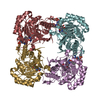
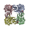
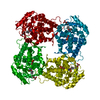
| Title | Structure of human guanosine monophosphate reductase GMPR1 in complex with GMP | ||||||
 Components Components | GMP REDUCTASE I | ||||||
 Keywords Keywords | OXIDOREDUCTASE / NUCLEOTIDE PATHWAY / TIM BARREL | ||||||
| Function / homology |  Function and homology informationGMP reductase / GMP reductase complex / GMP reductase activity / purine nucleotide metabolic process / Purine salvage / purine nucleobase metabolic process / response to cold / metal ion binding / cytosol Function and homology informationGMP reductase / GMP reductase complex / GMP reductase activity / purine nucleotide metabolic process / Purine salvage / purine nucleobase metabolic process / response to cold / metal ion binding / cytosolSimilarity search - Function | ||||||
| Biological species |  HOMO SAPIENS (human) HOMO SAPIENS (human) | ||||||
| Method | X-RAY DIFFRACTION / MOLECULAR REPLACEMENT / Resolution: 2.4 Å | ||||||
 Authors Authors | Bunkoczi, G. / Haroniti, A. / Ng, S. / von Delft, F. / Gileadi, O. / Oppermann, U. / Arrowsmith, C. / Edwards, A. / Sundstrom, M. | ||||||
 Citation Citation | Journal: To be Published Title: Structure of Human Guanosine Monophosphate Reductase Gmpr1 in Complex with Gmp Authors: Bunkoczi, G. / Haroniti, A. / Ng, S. / von Delft, F. / Gileadi, O. / Oppermann, U. / Arrowsmith, C. / Edwards, A. / Sundstrom, M. | ||||||
| History |
| ||||||
| Remark 700 | SHEET DETERMINATION METHOD: DSSP THE SHEETS PRESENTED AS "AC BB" IN EACH CHAIN ON SHEET RECORDS ... SHEET DETERMINATION METHOD: DSSP THE SHEETS PRESENTED AS "AC BB" IN EACH CHAIN ON SHEET RECORDS BELOW ARE ACTUALLY 9-STRANDED BARRELS REPRESENTED BY 10-STRANDED SHEETS IN WHICH THE FIRST AND LAST STRANDS ARE IDENTICAL. THE SHEETS PRESENTED AS "CB DA" IN EACH CHAIN ON SHEET RECORDS BELOW ARE ACTUALLY 8-STRANDED BARRELS REPRESENTED BY 9-STRANDED SHEETS IN WHICH THE FIRST AND LAST STRANDS ARE IDENTICAL. |
- Structure visualization
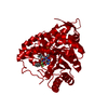
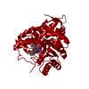
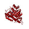
| Structure viewer | Molecule: MolmilJmol/JSmol |
|---|
- Downloads & links
-Download
| PDBx/mmCIF format | 2bwg.cif.gz | 263.9 KB | Display | PDBx/mmCIF format |
|---|---|---|---|---|
| PDB format | pdb2bwg.ent.gz | 209.5 KB | Display | PDB format |
| PDBx/mmJSON format | 2bwg.json.gz | Tree view | PDBx/mmJSON format | |
| Others |  Other downloads Other downloads |
-Validation report
| Arichive directory | https://data.pdbj.org/pub/pdb/validation_reports/bw/2bwgftp://data.pdbj.org/pub/pdb/validation_reports/bw/2bwg | HTTPS FTP |
|---|
-Related structure data
| Related structure data |  2bleC  1eepS C: citing same article ( S: Starting model for refinement |
|---|---|
| Similar structure data |


-Links
 PDBj
PDBj

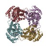
- Assembly
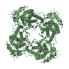
| Deposited unit | 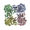
| |||||||||||||||||||||||||||||||||||||||||||||||||||||||||||||
|---|---|---|---|---|---|---|---|---|---|---|---|---|---|---|---|---|---|---|---|---|---|---|---|---|---|---|---|---|---|---|---|---|---|---|---|---|---|---|---|---|---|---|---|---|---|---|---|---|---|---|---|---|---|---|---|---|---|---|---|---|---|---|
| 1 |
| |||||||||||||||||||||||||||||||||||||||||||||||||||||||||||||
| Unit cell |
| |||||||||||||||||||||||||||||||||||||||||||||||||||||||||||||
| Noncrystallographic symmetry (NCS) | NCS domain:
NCS domain segments:
NCS oper:
|
-Components
| #1: Protein | / GUANOSINE 5'-MONOPHOSPHATE OXIDOREDUCTASE 1 / GUANOSINE MONOPHOSPHATE REDUCTASE 1 Mass: 39991.539 Da / Num. of mol.: 4 Source method: isolated from a genetically manipulated source Source: (gene. exp.) HOMO SAPIENS (human) / Plasmid: PNIC-BSA4 / Production host:  ESCHERICHIA COLI (E. coli) / Strain (production host): BL21(DE3) / References: UniProt: P36959, GMP reductase ESCHERICHIA COLI (E. coli) / Strain (production host): BL21(DE3) / References: UniProt: P36959, GMP reductase#2: Chemical | ChemComp-K /   Mass: 39.098 Da / Num. of mol.: 4 / Source method: obtained synthetically / Formula: K Mass: 39.098 Da / Num. of mol.: 4 / Source method: obtained synthetically / Formula: K#3: Chemical | ChemComp-5GP / Guanosine monophosphate  Mass: 363.221 Da / Num. of mol.: 4 / Source method: obtained synthetically / Formula: C10H14N5O8P Mass: 363.221 Da / Num. of mol.: 4 / Source method: obtained synthetically / Formula: C10H14N5O8P#4: Water | ChemComp-HOH / | Water Mass: 18.015 Da / Num. of mol.: 193 / Source method: isolated from a natural source / Formula: H2O Mass: 18.015 Da / Num. of mol.: 193 / Source method: isolated from a natural source / Formula: H2OSequence details | RESIDUES 1-22 CONSTITUTE A HIS-TAG THE SEQUENCE VARIANT AT RESIDUE 256 IS DESCRIBED BY THE UNIPROT ...RESIDUES 1-22 CONSTITUTE | |
|---|
-Experimental details
-Experiment
| Experiment | Method: X-RAY DIFFRACTION / Number of used crystals: 1 |
|---|
- Sample preparation
| Crystal | Density Matthews: 2.2 Å3/Da / Density % sol: 42.4 % |
|---|---|
| Crystal grow | Details: 16% PEG3350 0.30 M K3CIT |
-Data collection
| Diffraction | Mean temperature: 100 K |
|---|---|
| Diffraction source | Source: ROTATING ANODE / Type: RIGAKU FR-E / Wavelength: 1.5418 |
| Detector | Type: RIGAKU IMAGE PLATE / Detector: IMAGE PLATE / Date: Jan 31, 2005 |
| Radiation | Monochromator: OSMIC HR MULTILAYER OPTICS / Protocol: SINGLE WAVELENGTH / Monochromatic (M) / Laue (L): M / Scattering type: x-ray |
| Radiation wavelength | Wavelength: 1.5418 Å / Relative weight: 1 |
| Reflection | Resolution: 2.4→19.9 Å / Num. obs: 154258 / % possible obs: 90.1 % / Observed criterion σ(I): 0 / Redundancy: 2.83 % / Rmerge(I) obs: 0.12 / Net I/σ(I): 7.14 |
| Reflection shell | Resolution: 2.4→2.5 Å / Redundancy: 2.27 % / Rmerge(I) obs: 0.37 / Mean I/σ(I) obs: 2 / % possible all: 80.8 |
- Processing
| Software |
| ||||||||||||||||||||||||||||||||||||||||||||||||||||||||||||||||||||||||||||||||||||||||||||||||||||||||||||||||||||||||||||||||||||||||||||||||||||||||||||||||||||||||||||||||||||||
|---|---|---|---|---|---|---|---|---|---|---|---|---|---|---|---|---|---|---|---|---|---|---|---|---|---|---|---|---|---|---|---|---|---|---|---|---|---|---|---|---|---|---|---|---|---|---|---|---|---|---|---|---|---|---|---|---|---|---|---|---|---|---|---|---|---|---|---|---|---|---|---|---|---|---|---|---|---|---|---|---|---|---|---|---|---|---|---|---|---|---|---|---|---|---|---|---|---|---|---|---|---|---|---|---|---|---|---|---|---|---|---|---|---|---|---|---|---|---|---|---|---|---|---|---|---|---|---|---|---|---|---|---|---|---|---|---|---|---|---|---|---|---|---|---|---|---|---|---|---|---|---|---|---|---|---|---|---|---|---|---|---|---|---|---|---|---|---|---|---|---|---|---|---|---|---|---|---|---|---|---|---|---|---|
| Refinement | Method to determine structure: MOLECULAR REPLACEMENT Starting model: PDB ENTRY 1EEP Resolution: 2.4→40 Å / Cor.coef. Fo:Fc: 0.941 / Cor.coef. Fo:Fc free: 0.914 / SU B: 18.356 / SU ML: 0.222 / TLS residual ADP flag: UNVERIFIED / Cross valid method: THROUGHOUT / ESU R: 0.65 / ESU R Free: 0.29 / Stereochemistry target values: MAXIMUM LIKELIHOOD / Details: HYDROGENS HAVE BEEN ADDED IN THE RIDING POSITIONS.
| ||||||||||||||||||||||||||||||||||||||||||||||||||||||||||||||||||||||||||||||||||||||||||||||||||||||||||||||||||||||||||||||||||||||||||||||||||||||||||||||||||||||||||||||||||||||
| Solvent computation | Ion probe radii: 0.8 Å / Shrinkage radii: 0.8 Å / VDW probe radii: 1.2 Å / Solvent model: MASK | ||||||||||||||||||||||||||||||||||||||||||||||||||||||||||||||||||||||||||||||||||||||||||||||||||||||||||||||||||||||||||||||||||||||||||||||||||||||||||||||||||||||||||||||||||||||
| Displacement parameters | Biso mean: 19.81 Å2
| ||||||||||||||||||||||||||||||||||||||||||||||||||||||||||||||||||||||||||||||||||||||||||||||||||||||||||||||||||||||||||||||||||||||||||||||||||||||||||||||||||||||||||||||||||||||
| Refinement step | Cycle: LAST / Resolution: 2.4→40 Å
| ||||||||||||||||||||||||||||||||||||||||||||||||||||||||||||||||||||||||||||||||||||||||||||||||||||||||||||||||||||||||||||||||||||||||||||||||||||||||||||||||||||||||||||||||||||||
| Refine LS restraints |
|