 Movie
Movie Controller


+ Open data

- Basic information
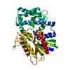
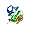
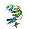
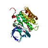
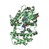
| Entry | Database: PDB / ID: 2bva | ||||||
|---|---|---|---|---|---|---|---|
| Title | Crystal structure of the human P21-activated kinase 4 | ||||||
 Components Components | P21-ACTIVATED KINASE 4 | ||||||
 Keywords Keywords |  TRANSFERASE / PROTEIN KINASE / STE20 / PAK4 / ATP-BINDING TRANSFERASE / PROTEIN KINASE / STE20 / PAK4 / ATP-BINDING | ||||||
| Function / homology |  Function and homology information Function and homology informationsignal transduction => GO:0007165 / dendritic spine development / cadherin binding involved in cell-cell adhesion / Activation of RAC1 / stress-activated protein kinase signaling cascade / RHOV GTPase cycle / activation of protein kinase activity / RHOJ GTPase cycle / RHOQ GTPase cycle / regulation of MAPK cascade ...signal transduction => GO:0007165 / dendritic spine development / cadherin binding involved in cell-cell adhesion / Activation of RAC1 / stress-activated protein kinase signaling cascade / RHOV GTPase cycle / activation of protein kinase activity / RHOJ GTPase cycle / RHOQ GTPase cycle / regulation of MAPK cascade / RHOH GTPase cycle / CDC42 GTPase cycle / RHOU GTPase cycle / cellular response to organic cyclic compound / RHOG GTPase cycle / RAC2 GTPase cycle / RAC3 GTPase cycle / negative regulation of endothelial cell apoptotic process / cytoskeleton organization / RAC1 GTPase cycle / regulation of cell growth / adherens junction / positive regulation of angiogenesis / cell migration / non-specific serine/threonine protein kinase / protein kinase activity / intracellular signal transduction / cell cycle / phosphorylation / protein serine kinase activity / focal adhesion / protein serine/threonine kinase activity / apoptotic process / Golgi apparatus / signal transduction / ATP binding / cytosol / cytoplasmSimilarity search - Function | ||||||
| Biological species |  HOMO SAPIENS (human) HOMO SAPIENS (human) | ||||||
| Method | X-RAY DIFFRACTION / SYNCHROTRON / MOLECULAR REPLACEMENT / Resolution: 2.3 Å | ||||||
 Authors Authors | Debreczeni, J.E. / Bunkoczi, G. / Eswaran, J. / Filippakopoulos, P. / Das, S. / Fedorov, O. / Sundstrom, M. / Arrowsmith, C. / Edwards, A. / von Delft, F. / Knapp, S. | ||||||
 Citation Citation | Journal: Structure / Year: 2007 Title: Crystal Structures of the P21-Activated Kinases Pak4, Pak5, and Pak6 Reveal Catalytic Domain Plasticity of Active Group II Paks. Authors: Eswaran, J. / Lee, W.H. / Debreczeni, J.E. / Filippakopoulos, P. / Turnbull, A. / Fedorov, O. / Deacon, S.W. / Peterson, J.R. / Knapp, S. | ||||||
| History |
|
- Structure visualization
| Structure viewer | Molecule: MolmilJmol/JSmol |
|---|
- Downloads & links
-Download
| PDBx/mmCIF format | 2bva.cif.gz | 117.4 KB | Display | PDBx/mmCIF format |
|---|---|---|---|---|
| PDB format | pdb2bva.ent.gz | 90.8 KB | Display | PDB format |
| PDBx/mmJSON format | 2bva.json.gz | Tree view | PDBx/mmJSON format | |
| Others |  Other downloads Other downloads |
-Validation report
| Arichive directory | https://data.pdbj.org/pub/pdb/validation_reports/bv/2bvaftp://data.pdbj.org/pub/pdb/validation_reports/bv/2bva | HTTPS FTP |
|---|
-Related structure data
| Related structure data |  2c30C 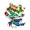 2cdzC 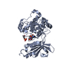 2f57C 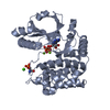 1u5rS S: Starting model for refinement C: citing same article ( |
|---|---|
| Similar structure data |


-Links
 PDBj
PDBj


- Assembly
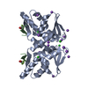
| Deposited unit | 
| ||||||||
|---|---|---|---|---|---|---|---|---|---|
| 1 | 
| ||||||||
| 2 | 
| ||||||||
| Unit cell |
| ||||||||
| Noncrystallographic symmetry (NCS) | NCS oper: (Code: given Matrix: (-0.49972, -0.86616, -0.00676), Vector : |
-Components
| #1: Protein | Mass: 33011.371 Da / Num. of mol.: 2 / Fragment: KINASE DOMAIN, RESIDUES 300-591 Source method: isolated from a genetically manipulated source Details: PHOSPHORYLATION ON SER 474 A AND SER 474 B / Source: (gene. exp.) HOMO SAPIENS (human) / Plasmid: PGEX6B-C001 / Production host:  ESCHERICHIA COLI (E. coli) / Strain (production host): BL21(DE3) / Variant (production host): R (PHAGE RESISTANT) ESCHERICHIA COLI (E. coli) / Strain (production host): BL21(DE3) / Variant (production host): R (PHAGE RESISTANT)References: UniProt: Q8NCH5, UniProt: O96013*PLUS, EC: 2.7.1.37 #2: Water | ChemComp-HOH / | Water Mass: 18.015 Da / Num. of mol.: 36 / Source method: isolated from a natural source / Formula: H2O Mass: 18.015 Da / Num. of mol.: 36 / Source method: isolated from a natural source / Formula: H2O |
|---|
-Experimental details
-Experiment
| Experiment | Method: X-RAY DIFFRACTION / Number of used crystals: 1 |
|---|
- Sample preparation
| Crystal | Density Matthews: 3 Å3/Da / Density % sol: 59 % |
|---|---|
| Crystal grow | Method: vapor diffusion, sitting drop / Details: SITTING DROPS, 1.5 M NACL, 10% ETHANOL |
-Data collection
| Diffraction | Mean temperature: 100 K |
|---|---|
| Diffraction source | Source: SYNCHROTRON / Site: SLS  / Beamline: X10SA / Wavelength: 0.968 / Beamline: X10SA / Wavelength: 0.968 |
| Detector | Type: MARRESEARCH / Detector: CCD / Date: Apr 23, 2005 |
| Radiation | Protocol: SINGLE WAVELENGTH / Monochromatic (M) / Laue (L): M / Scattering type: x-ray |
| Radiation wavelength | Wavelength: 0.968 Å / Relative weight: 1 |
| Reflection | Resolution: 2.3→40.4 Å / Num. obs: 38417 / % possible obs: 99.8 % / Observed criterion σ(I): 3.5 / Redundancy: 2.7 % / Rmerge(I) obs: 0.06 / Net I/σ(I): 11.86 |
| Reflection shell | Resolution: 2.3→2.4 Å / Redundancy: 2.7 % / Rmerge(I) obs: 0.28 / Mean I/σ(I) obs: 3.55 / % possible all: 100 |
- Processing
| Software |
| |||||||||||||||||||||||||||||||||
|---|---|---|---|---|---|---|---|---|---|---|---|---|---|---|---|---|---|---|---|---|---|---|---|---|---|---|---|---|---|---|---|---|---|---|
| Refinement | Method to determine structure: MOLECULAR REPLACEMENT Starting model: PDB ENTRY 1U5R Resolution: 2.3→20 Å / Num. parameters: 17176 / Num. restraintsaints: 22479 / Cross valid method: FREE R-VALUE / σ(F): 0 Stereochemistry target values: ENGH AND HUBER BASED ON HIC-UP ENTRY Details: 1. HYDROGENS WERE ADDED ON RIDING POSITIONS 2. TWIN REFINEMENT: PERFECT MEROHEDRAL TWIN, APPARENT SPACEGROUP P3221, REAL SPACEGROUP P32, TWIN LAW: 010 100 00-1, BASF: 0.5006 3. NCS RESTRAINTS WERE USED
| |||||||||||||||||||||||||||||||||
| Refine analyze | Num. disordered residues: 0 / Occupancy sum hydrogen: 13065 / Occupancy sum non hydrogen: 12879 | |||||||||||||||||||||||||||||||||
| Refinement step | Cycle: LAST / Resolution: 2.3→20 Å
| |||||||||||||||||||||||||||||||||
| Refine LS restraints |
|
