 Movie
Movie Controller

[English] 日本語
 Yorodumi
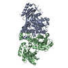
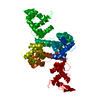
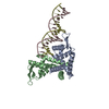
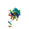
- PDB-2bsf: Structure of the C-terminal receptor-binding domain of avian reov... -
+ Open data

- Basic information
| Entry | Database: PDB / ID: 2bsf | ||||||
|---|---|---|---|---|---|---|---|
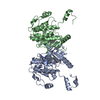
| Title | Structure of the C-terminal receptor-binding domain of avian reovirus fibre sigmaC, Zn crystal form. | ||||||
 Components Components | SIGMA C CAPSID PROTEIN | ||||||
 Keywords Keywords |  VIRAL PROTEIN / ORTHOREOVIRUS / TRIPLE BETA-SPIRAL / BETA-BARREL VIRAL PROTEIN / ORTHOREOVIRUS / TRIPLE BETA-SPIRAL / BETA-BARREL | ||||||
| Function / homology |  Function and homology informationviral capsid / symbiont entry into host cell / virion attachment to host cell Function and homology informationviral capsid / symbiont entry into host cell / virion attachment to host cellSimilarity search - Function | ||||||
| Biological species |  AVIAN REOVIRUS AVIAN REOVIRUS | ||||||
| Method | X-RAY DIFFRACTION / SYNCHROTRON / MAD / Resolution: 2.1 Å | ||||||
 Authors Authors | Guardado Calvo, P. / Fox, G.C. / Hermo Parrado, X.L. / Llamas-Saiz, A.L. / van Raaij, M.J. | ||||||
 Citation Citation | Journal: J.Mol.Biol. / Year: 2005 Title: Structure of the Carboxy-Terminal Receptor-Binding Domain of Avian Reovirus Fibre Sigmac Authors: Guardado Calvo, P. / Fox, G.C. / Hermo Parrado, X.L. / Llamas-Saiz, A.L. / Costas, C. / Martinez-Costas, J. / Benavente, J. / van Raaij, M.J. #1: Journal: Acta Crystallogr.,Sect.F / Year: 2005 Title: Crystallisation of the C-Terminal Globular Domain of Avian Reovirus Fibre Authors: van Raaij, M.J. / Hermo Parrado, X.L. / Guardado Calvo, P. / Fox, G.C. / Llamas-Saiz, A.L. / Costas, C. / Martinez-Costas, J. / Benavente, J. #2: Journal: Embo J. / Year: 2002Title: Crystal Structure of Reovirus Attachment Protein Sigma1 Reveals Evolutionary Relationship to Adenovirus Fiber Authors: Chappell, J.D. / Prota, A.E. / Dermody, T.S. / Stehle, T. #3: Journal: J.Virol. / Year: 1997 Title: Protein Architecture of Avian Reovirus S1133 and Identification of the Cell Attachment Protein Authors: Martinez-Costas, J. / Grande, A. / Varela, R. / Garcia-Martinez, C. / Benavente, J. #4: Journal: Virology / Year: 2000 Title: Oligomerization and Cell-Binding Properties of the Avian Reovirus Cell-Attachment Protein Sigmac Authors: Grande, A. / Rodriguez, E. / Costas, C. / Everitt, E. / Benavente, J. #5: Journal: Virology / Year: 2001 Title: The Avian Reovirus Genome Segment S1 is a Functionally Tricistronic Gene that Expresses One Structure and Two Nonstructural Proteins in Infected Cells Authors: Bodelon, G. / Labrada, L. / Martinez-Costas, J. / Benavente, J. #6: Journal: J.Gen.Virol. / Year: 2002 Title: Subunit Composition and Conformational Stability of the Oligomeric Form of the Avian Reovirus Cell-Attachment Protein Sigmac Authors: Grande, A. / Costas, C. / Benavente, J. | ||||||
| History |
| ||||||
| Remark 700 | SHEET THE SHEET STRUCTURE OF THIS MOLECULE IS BIFURCATED. IN ORDER TO REPRESENT THIS FEATURE IN ... SHEET THE SHEET STRUCTURE OF THIS MOLECULE IS BIFURCATED. IN ORDER TO REPRESENT THIS FEATURE IN THE SHEET RECORDS BELOW, TWO SHEETS ARE DEFINED. |
- Structure visualization
| Structure viewer | Molecule: MolmilJmol/JSmol |
|---|
- Downloads & links
-Download
| PDBx/mmCIF format | 2bsf.cif.gz | 51.6 KB | Display | PDBx/mmCIF format |
|---|---|---|---|---|
| PDB format | pdb2bsf.ent.gz | 36.9 KB | Display | PDB format |
| PDBx/mmJSON format | 2bsf.json.gz | Tree view | PDBx/mmJSON format | |
| Others |  Other downloads Other downloads |
-Validation report
| Arichive directory | https://data.pdbj.org/pub/pdb/validation_reports/bs/2bsfftp://data.pdbj.org/pub/pdb/validation_reports/bs/2bsf | HTTPS FTP |
|---|
-Related structure data


-Links
 PDBj
PDBj- Assembly
| Deposited unit | 
| ||||||||
|---|---|---|---|---|---|---|---|---|---|
| 1 | 
| ||||||||
| Unit cell |
| ||||||||
| Components on special symmetry positions |
|
-Components
| #1: Protein | Mass: 18912.947 Da / Num. of mol.: 1 Fragment: C-TERMINAL RECEPTOR-BINDING REGION, RESIDUES 151-326 Source method: isolated from a genetically manipulated source Source: (gene. exp.) AVIAN REOVIRUS / Strain: S1133Description: THE AVIAN REOVIRUS STRAIN S1133 WAS ORIGINALLY PROVIDED BY DR. PHILIP I.MARCUS WHEN DR. J. BENAVENTE WAS A ROCHE VISITING SCIENTIST IN THE LABORATORY OF DR. A.SHATKIN Plasmid: PET28CPLUS / Production host:  ESCHERICHIA COLI BL21(DE3) (bacteria) / References: UniProt: O12287, UniProt: Q992I2*PLUS ESCHERICHIA COLI BL21(DE3) (bacteria) / References: UniProt: O12287, UniProt: Q992I2*PLUS | ||||
|---|---|---|---|---|---|
| #2: Chemical | Sulfate  Mass: 96.063 Da / Num. of mol.: 3 / Source method: obtained synthetically / Formula: SO4 Mass: 96.063 Da / Num. of mol.: 3 / Source method: obtained synthetically / Formula: SO4#3: Chemical | ChemComp-ZN / |   Mass: 65.409 Da / Num. of mol.: 1 / Source method: obtained synthetically / Formula: Zn Mass: 65.409 Da / Num. of mol.: 1 / Source method: obtained synthetically / Formula: Zn#4: Water | ChemComp-HOH / | Water Mass: 18.015 Da / Num. of mol.: 164 / Source method: isolated from a natural source / Formula: H2O Mass: 18.015 Da / Num. of mol.: 164 / Source method: isolated from a natural source / Formula: H2O |
-Experimental details
-Experiment
| Experiment | Method: X-RAY DIFFRACTION / Number of used crystals: 1 |
|---|
- Sample preparation
| Crystal | Density Matthews: 3.2 Å3/Da / Density % sol: 0.61 % |
|---|---|
| Crystal grow | pH: 8.5 Details: 100 MM TRIS-HCL PH 8.5, 1.5 M AMMONIUM SULPHATE, 12% GLYCEROL, 10 MM ZINC SULPHATE |
-Data collection
| Diffraction | Mean temperature: 100 K | |||||||||||||||
|---|---|---|---|---|---|---|---|---|---|---|---|---|---|---|---|---|
| Diffraction source | Source: SYNCHROTRON / Site: ESRF  / Beamline: BM16 / Wavelength: 1.28191,1.15872, 1.28271, 1.28194 / Beamline: BM16 / Wavelength: 1.28191,1.15872, 1.28271, 1.28194 | |||||||||||||||
| Detector | Type: MARRESEARCH / Detector: CCD / Date: Dec 17, 2004 / Details: TOROIDAL MIRROR | |||||||||||||||
| Radiation | Monochromator: DOUBLE CRYSTAL / Protocol: SINGLE WAVELENGTH / Monochromatic (M) / Laue (L): M / Scattering type: x-ray | |||||||||||||||
| Radiation wavelength |
| |||||||||||||||
| Reflection | Resolution: 2.1→30 Å / Num. obs: 14231 / % possible obs: 98.7 % / Observed criterion σ(I): 0 / Redundancy: 7.8 % / Biso Wilson estimate: 28.27 Å2 / Rmerge(I) obs: 0.07 / Net I/σ(I): 7.3 | |||||||||||||||
| Reflection shell | Resolution: 2.1→2.21 Å / Redundancy: 8 % / Rmerge(I) obs: 0.29 / Mean I/σ(I) obs: 2.6 / % possible all: 97.7 |
- Processing
| Software |
| ||||||||||||||||||||||||||||||||||||||||||||||||||||||||||||||||||||||||||||||||||||||||||||||||||||||||||||||||||||||||||||||||||||||||||||||||||||||||||||||||||||||||||||||||||||||
|---|---|---|---|---|---|---|---|---|---|---|---|---|---|---|---|---|---|---|---|---|---|---|---|---|---|---|---|---|---|---|---|---|---|---|---|---|---|---|---|---|---|---|---|---|---|---|---|---|---|---|---|---|---|---|---|---|---|---|---|---|---|---|---|---|---|---|---|---|---|---|---|---|---|---|---|---|---|---|---|---|---|---|---|---|---|---|---|---|---|---|---|---|---|---|---|---|---|---|---|---|---|---|---|---|---|---|---|---|---|---|---|---|---|---|---|---|---|---|---|---|---|---|---|---|---|---|---|---|---|---|---|---|---|---|---|---|---|---|---|---|---|---|---|---|---|---|---|---|---|---|---|---|---|---|---|---|---|---|---|---|---|---|---|---|---|---|---|---|---|---|---|---|---|---|---|---|---|---|---|---|---|---|---|
| Refinement | Method to determine structure: MAD / Resolution: 2.1→74.54 Å / Cor.coef. Fo:Fc: 0.961 / Cor.coef. Fo:Fc free: 0.927 / SU B: 3.41 / SU ML: 0.093 / Cross valid method: THROUGHOUT / ESU R: 0.166 / ESU R Free: 0.167 / Stereochemistry target values: MAXIMUM LIKELIHOOD / Details: HYDROGENS HAVE BEEN ADDED IN THE RIDING POSITIONS.
| ||||||||||||||||||||||||||||||||||||||||||||||||||||||||||||||||||||||||||||||||||||||||||||||||||||||||||||||||||||||||||||||||||||||||||||||||||||||||||||||||||||||||||||||||||||||
| Solvent computation | Ion probe radii: 0.8 Å / Shrinkage radii: 0.8 Å / VDW probe radii: 1.2 Å / Solvent model: MASK | ||||||||||||||||||||||||||||||||||||||||||||||||||||||||||||||||||||||||||||||||||||||||||||||||||||||||||||||||||||||||||||||||||||||||||||||||||||||||||||||||||||||||||||||||||||||
| Displacement parameters | Biso mean: 25.93 Å2
| ||||||||||||||||||||||||||||||||||||||||||||||||||||||||||||||||||||||||||||||||||||||||||||||||||||||||||||||||||||||||||||||||||||||||||||||||||||||||||||||||||||||||||||||||||||||
| Refinement step | Cycle: LAST / Resolution: 2.1→74.54 Å
| ||||||||||||||||||||||||||||||||||||||||||||||||||||||||||||||||||||||||||||||||||||||||||||||||||||||||||||||||||||||||||||||||||||||||||||||||||||||||||||||||||||||||||||||||||||||
| Refine LS restraints |
|