 Movie
Movie Controller

[English] 日本語
 Yorodumi
- PDB-2bov: Molecular recognition of an ADP-ribosylating Clostridium botulinu... -
+ Open data

- Basic information
| Entry | Database: PDB / ID: 2bov | |||||||||
|---|---|---|---|---|---|---|---|---|---|---|
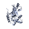
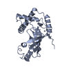
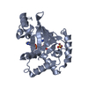
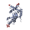
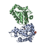
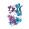
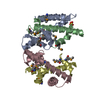
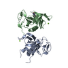
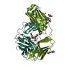
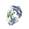
| Title | Molecular recognition of an ADP-ribosylating Clostridium botulinum C3 exoenzyme by RalA GTPase | |||||||||
 Components Components |
| |||||||||
 Keywords Keywords |  TRANSFERASE / C3BOT / EXOENZYME / RALA / GTPASE / ADP / RIBOSYLATING TOXIN / GTP-BINDING / LIPOPROTEIN / PRENYLATION / GLYCOSYLTRANSFERASE / NAD TRANSFERASE / C3BOT / EXOENZYME / RALA / GTPASE / ADP / RIBOSYLATING TOXIN / GTP-BINDING / LIPOPROTEIN / PRENYLATION / GLYCOSYLTRANSFERASE / NAD | |||||||||
| Function / homology |  Function and homology information Function and homology informationmembrane raft localization / Edg-2 lysophosphatidic acid receptor binding / establishment of protein localization to mitochondrion / regulation of exocytosis / Flemming body / regulation of postsynaptic neurotransmitter receptor internalization / positive regulation of filopodium assembly / positive regulation of epidermal growth factor receptor signaling pathway / positive regulation of mitochondrial fission / myosin binding ...membrane raft localization / Edg-2 lysophosphatidic acid receptor binding / establishment of protein localization to mitochondrion / regulation of exocytosis / Flemming body / regulation of postsynaptic neurotransmitter receptor internalization / positive regulation of filopodium assembly / positive regulation of epidermal growth factor receptor signaling pathway / positive regulation of mitochondrial fission / myosin binding / exocytosis / NAD+-protein ADP-ribosyltransferase activity / cleavage furrow / Transferases; Glycosyltransferases; Pentosyltransferases / p38MAPK events / Gene and protein expression by JAK-STAT signaling after Interleukin-12 stimulation / nucleotidyltransferase activity / small monomeric GTPase / G protein activity / synaptic membrane / neural tube closure / Translocation of SLC2A4 (GLUT4) to the plasma membrane / regulation of actin cytoskeleton organization / Schaffer collateral - CA1 synapse / cytoplasmic vesicle membrane / receptor internalization / GDP binding / chemotaxis / ATPase binding / Ras protein signal transduction / cell cycle / cell division / focal adhesion / GTPase activity / ubiquitin protein ligase binding / GTP binding / cell surface / signal transduction / mitochondrion / extracellular exosome / extracellular region / plasma membraneSimilarity search - Function | |||||||||
| Biological species |  HOMO SAPIENS (human) HOMO SAPIENS (human)  CLOSTRIDIUM BOTULINUM (bacteria) CLOSTRIDIUM BOTULINUM (bacteria) | |||||||||
| Method | X-RAY DIFFRACTION / SYNCHROTRON / MOLECULAR REPLACEMENT / Resolution: 2.66 Å | |||||||||
 Authors Authors | Holbourn, K.P. / Sutton, J.M. / Evans, H.R. / Shone, C.C. / Acharya, K.R. | |||||||||
 Citation Citation | Journal: Proc.Natl.Acad.Sci.USA / Year: 2005 Title: Molecular Recognition of an Adp-Ribosylating Clostridium Botulinum C3 Exoenzyme by Rala Gtpase Authors: Holbourn, K.P. / Sutton, J.M. / Evans, H.R. / Shone, C.C. / Acharya, K.R. | |||||||||
| History |
|
- Structure visualization
| Structure viewer | Molecule: MolmilJmol/JSmol |
|---|
- Downloads & links
-Download
| PDBx/mmCIF format | 2bov.cif.gz | 99.7 KB | Display | PDBx/mmCIF format |
|---|---|---|---|---|
| PDB format | pdb2bov.ent.gz | 73 KB | Display | PDB format |
| PDBx/mmJSON format | 2bov.json.gz | Tree view | PDBx/mmJSON format | |
| Others |  Other downloads Other downloads |
-Validation report
| Arichive directory | https://data.pdbj.org/pub/pdb/validation_reports/bo/2bovftp://data.pdbj.org/pub/pdb/validation_reports/bo/2bov | HTTPS FTP |
|---|
-Related structure data


-Links
 PDBj
PDBj


- Assembly
| Deposited unit | 
| ||||||||
|---|---|---|---|---|---|---|---|---|---|
| 1 |
| ||||||||
| Unit cell |
|
-Components
| #1: Protein | Mass: 23603.846 Da / Num. of mol.: 1 Source method: isolated from a genetically manipulated source Source: (gene. exp.) HOMO SAPIENS (human) / Plasmid: PGEX-2T / Production host: ESCHERICHIA COLI (E. coli) / Strain (production host): JM109 / References: UniProt: P11233 |
|---|---|
| #2: Protein | Mass: 27877.172 Da / Num. of mol.: 1 Source method: isolated from a genetically manipulated source Source: (gene. exp.) CLOSTRIDIUM BOTULINUM (bacteria) / Plasmid: PMAL / Production host: ESCHERICHIA COLI (E. coli) / Strain (production host): JM109References: UniProt: P15879, Transferases; Glycosyltransferases; Pentosyltransferases |
| #3: Chemical | ChemComp-GDP / Guanosine diphosphate  Type: RNA linking / Mass: 443.201 Da / Num. of mol.: 1 / Source method: obtained synthetically / Formula: C10H15N5O11P2 / Comment: GDP, energy-carrying molecule*YM Type: RNA linking / Mass: 443.201 Da / Num. of mol.: 1 / Source method: obtained synthetically / Formula: C10H15N5O11P2 / Comment: GDP, energy-carrying molecule*YM |
| #4: Chemical | ChemComp-MG /   Mass: 24.305 Da / Num. of mol.: 1 / Source method: obtained synthetically / Formula: Mg Mass: 24.305 Da / Num. of mol.: 1 / Source method: obtained synthetically / Formula: Mg |
| #5: Water | ChemComp-HOH / Water Mass: 18.015 Da / Num. of mol.: 261 / Source method: isolated from a natural source / Formula: H2O Mass: 18.015 Da / Num. of mol.: 261 / Source method: isolated from a natural source / Formula: H2O |
| Compound details | FUNCTION: ADP-RIBOSYLATI |
-Experimental details
-Experiment
| Experiment | Method: X-RAY DIFFRACTION / Number of used crystals: 1 |
|---|
- Sample preparation
| Crystal | Density Matthews: 2.7 Å3/Da / Density % sol: 54 % |
|---|---|
| Crystal grow | pH: 7.3 Details: 0.1M HEPES PH 7.3 12-15% ETHYLENE GLYCOL 14-18% PEG 8000 |
-Data collection
| Diffraction | Mean temperature: 100 K |
|---|---|
| Diffraction source | Source: SYNCHROTRON / Site: SRS  / Beamline: PX9.6 / Wavelength: 0.87 / Beamline: PX9.6 / Wavelength: 0.87 |
| Detector | Type: ADSC CCD / Detector: CCD / Date: Feb 22, 2004 |
| Radiation | Protocol: SINGLE WAVELENGTH / Monochromatic (M) / Laue (L): M / Scattering type: x-ray |
| Radiation wavelength | Wavelength: 0.87 Å / Relative weight: 1 |
| Reflection | Resolution: 2.66→50 Å / Num. obs: 15556 / % possible obs: 99.8 % / Observed criterion σ(I): 2 / Redundancy: 15.6 % / Biso Wilson estimate: 27.2 Å2 / Rmerge(I) obs: 0.11 / Net I/σ(I): 14.6 |
| Reflection shell | Resolution: 2.66→2.76 Å / Rmerge(I) obs: 0.26 / Mean I/σ(I) obs: 5.8 / % possible all: 99.9 |
- Processing
| Software |
| ||||||||||||||||||||||||||||||||||||||||||||||||||||||||||||
|---|---|---|---|---|---|---|---|---|---|---|---|---|---|---|---|---|---|---|---|---|---|---|---|---|---|---|---|---|---|---|---|---|---|---|---|---|---|---|---|---|---|---|---|---|---|---|---|---|---|---|---|---|---|---|---|---|---|---|---|---|---|
| Refinement | Method to determine structure: MOLECULAR REPLACEMENT Starting model: PDB ENTRY 1G24 AND 1UAD Resolution: 2.66→50 Å / Rfactor Rfree error: 0.009 / Cross valid method: THROUGHOUT / σ(F): 0
| ||||||||||||||||||||||||||||||||||||||||||||||||||||||||||||
| Solvent computation | Solvent model: FLAT MODEL / Bsol: 40.3303 Å2 / ksol: 0.375286 e/Å3 | ||||||||||||||||||||||||||||||||||||||||||||||||||||||||||||
| Displacement parameters | Biso mean: 22.5 Å2
| ||||||||||||||||||||||||||||||||||||||||||||||||||||||||||||
| Refine analyze |
| ||||||||||||||||||||||||||||||||||||||||||||||||||||||||||||
| Refinement step | Cycle: LAST / Resolution: 2.66→50 Å
| ||||||||||||||||||||||||||||||||||||||||||||||||||||||||||||
| Refine LS restraints |
| ||||||||||||||||||||||||||||||||||||||||||||||||||||||||||||
| LS refinement shell | Resolution: 2.66→2.83 Å / Rfactor Rfree error: 0.025 / Total num. of bins used: 6
|
