Mass: 18.015 Da / Num. of mol.: 249 / Source method: isolated from a natural source / Formula: H2O
Compound details
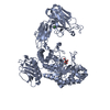
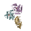
BINDS TO GTP-BOUND FORM OF RHO AND TO PROFILIN. ACTS IN A RHO-DEPENDENT MANNER TO RECRUIT PROFILIN ...BINDS TO GTP-BOUND FORM OF RHO AND TO PROFILIN. ACTS IN A RHO-DEPENDENT MANNER TO RECRUIT PROFILIN TO THE MEMBRANE, WHERE IT PROMOTES ACTIN POLYMERIZATION. IT IS REQUIRED FOR CYTOKINESIS, STRESS FIBER FORMATION, AND TRANSCRIPTIONAL ACTIVATION OF THE SERUM RESPONSE FACTOR.
-
Experimental details
-
Experiment
Experiment
Method: X-RAY DIFFRACTION / Number of used crystals: 1
-
Sample preparation
Crystal
Density Matthews: 4.5 Å3/Da / Density % sol: 72.9 %
Crystal grow
Method: vapor diffusion, hanging drop / pH: 7.25 Details: HANGING DROP VAPOR DIFFUSION; PROTEIN: 11 MG/ML IN 100 MM HEPES, PH 7.25; RESERVOIR: 100 MM HEPES, PH 7.25, 9% PEG3350
Protocol: SINGLE WAVELENGTH / Monochromatic (M) / Laue (L): M / Scattering type: x-ray
Radiation wavelength
Wavelength: 1.00691 Å / Relative weight: 1
Reflection
Resolution: 2.4→46 Å / Num. obs: 61321 / % possible obs: 99.8 % / Observed criterion σ(I): -3 / Redundancy: 5.9 % / Biso Wilson estimate: 55.09 Å2 / Rmerge(I) obs: 0.06 / Net I/σ(I): 27.3
Reflection shell
Resolution: 2.4→2.44 Å / Redundancy: 4.5 % / Rmerge(I) obs: 0.57 / Mean I/σ(I) obs: 2.1 / % possible all: 97.6
-
Processing
Software
Name
Version
Classification
REFMAC
5.2.0003
refinement
HKL-2000
datareduction
HKL-2000
datascaling
CNS
phasing
Refinement
Method to determine structure: SAD / Resolution: 2.4→20 Å / Cor.coef. Fo:Fc: 0.958 / Cor.coef. Fo:Fc free: 0.939 / SU B: 11.41 / SU ML: 0.136 / TLS residual ADP flag: UNVERIFIED / Cross valid method: THROUGHOUT / ESU R: 0.186 / ESU R Free: 0.176 / Stereochemistry target values: MAXIMUM LIKELIHOOD Details: HYDROGENS HAVE BEEN ADDED IN THE RIDING POSITIONS. RESIDUES 470 THROUGH 474 IN CHAIN A WERE MODELED AS ALANINES. BECAUSE SEQUENCE ASSIGNMENT WAS AMBIGUOUS DUE TO THE WEAK ELECTRON DENSITY IN THIS REGION
Rfactor
Num. reflection
% reflection
Selection details
Rfree
0.236
1542
2.5 %
RANDOM
Rwork
0.196
-
-
-
obs
0.197
59657
99.8 %
-
Solvent computation
Ion probe radii: 0.8 Å / Shrinkage radii: 0.8 Å / VDW probe radii: 1.2 Å / Solvent model: MASK
 Movie
Movie Controller

 Yorodumi
 Open data
 Basic information
 Components
 Transparency and translucency
  Keywords
 Function and homology information
 Authors
 Citation
 Structure visualization
 Downloads & links
 Other downloads
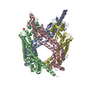


 PDBj
PDBj


 Assembly


 Mass: 35.453 Da / Num. of mol.: 1 / Source method: obtained synthetically / Formula: Cl
 Mass: 18.015 Da / Num. of mol.: 249 / Source method: isolated from a natural source / Formula: H2O
 Sample preparation
 / Beamline: 19-ID / Wavelength: 1.00691
  Processing