 Movie
Movie Controller

[English] 日本語
 Yorodumi
- PDB-2bmw: Ferredoxin: NADP+ Reductase Mutant With Thr 155 Replaced By Gly, ... -
+ Open data

- Basic information
| Entry | Database: PDB / ID: 2bmw | ||||||
|---|---|---|---|---|---|---|---|
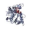
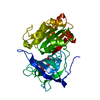
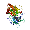
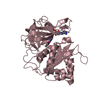
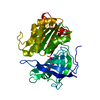
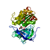
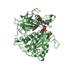
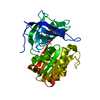
| Title | Ferredoxin: NADP+ Reductase Mutant With Thr 155 Replaced By Gly, Ala 160 Replaced By Thr, Leu 263 Replaced By Pro, Arg 264 Replaced By Pro and Gly 265 Replaced by Pro (T155G-A160T-L263P-R264P-G265P) | ||||||
 Components Components | FERREDOXIN--NADP REDUCTASE | ||||||
 Keywords Keywords |  OXIDOREDUCTASE / FLAVOPROTEIN / NADP / FAD / FNR / REDUCTASE REDUCTASE / PHYCOBILISOME / THYLAKOID OXIDOREDUCTASE / FLAVOPROTEIN / NADP / FAD / FNR / REDUCTASE REDUCTASE / PHYCOBILISOME / THYLAKOID | ||||||
| Function / homology |  Function and homology informationferredoxin-NADP+ reductase / ferredoxin-NADP+ reductase activity / phycobilisome / plasma membrane-derived thylakoid membrane / electron transport chain / NADP binding / flavin adenine dinucleotide binding Function and homology informationferredoxin-NADP+ reductase / ferredoxin-NADP+ reductase activity / phycobilisome / plasma membrane-derived thylakoid membrane / electron transport chain / NADP binding / flavin adenine dinucleotide bindingSimilarity search - Function | ||||||
| Biological species |  ANABAENA SP. (bacteria) ANABAENA SP. (bacteria) | ||||||
| Method | X-RAY DIFFRACTION / SYNCHROTRON / MOLECULAR REPLACEMENT / Resolution: 1.5 Å | ||||||
 Authors Authors | Martinez-Julvez, M. / Hermoso, J.A. / Perez-Dorado, I. / Medina, M. / Tejero, J. / Gomez-Moreno, C. | ||||||
 Citation Citation | Journal: Biochemistry / Year: 2009 Title: Protein Motifs Involved in Coenzyme Interaction and Enzymatic Efficiency in Anabaena Ferredoxin-Nadp+ Reductase. Authors: Peregrina, J.R. / Herguedas, B. / Hermoso, J.A. / Martinez-Julvez, M. / Medina, M. #1: Journal: Biophys.Chem. / Year: 2005 Title: Towards a New Therapeutic Target: Helicobacter Pylori Flavodoxin Authors: Cremades, N. / Bueno, M. / Toja, M. / Sancho, J. | ||||||
| History |
|
- Structure visualization
| Structure viewer | Molecule: MolmilJmol/JSmol |
|---|
- Downloads & links
-Download
| PDBx/mmCIF format | 2bmw.cif.gz | 88.4 KB | Display | PDBx/mmCIF format |
|---|---|---|---|---|
| PDB format | pdb2bmw.ent.gz | 64.9 KB | Display | PDB format |
| PDBx/mmJSON format | 2bmw.json.gz | Tree view | PDBx/mmJSON format | |
| Others |  Other downloads Other downloads |
-Validation report
| Arichive directory | https://data.pdbj.org/pub/pdb/validation_reports/bm/2bmwftp://data.pdbj.org/pub/pdb/validation_reports/bm/2bmw | HTTPS FTP |
|---|
-Related structure data
| Related structure data |  2vyqC 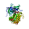 2vzlC 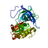 1queS S: Starting model for refinement C: citing same article ( |
|---|---|
| Similar structure data |


-Links
 PDBj
PDBj

- Assembly
| Deposited unit | 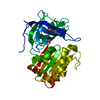
| ||||||||
|---|---|---|---|---|---|---|---|---|---|
| 1 |
| ||||||||
| Unit cell |
|
-Components
| #1: Protein | Mass: 34122.781 Da / Num. of mol.: 1 / Fragment: RESIDUES 137-440 / Mutation: YES Source method: isolated from a genetically manipulated source Details: MUTATIONS T155G, A160T, L263P, R264P AND G265P / Source: (gene. exp.) ANABAENA SP. (bacteria) / Strain: PCC 7119 / Production host: ESCHERICHIA COLI (E. coli) / References: UniProt: P21890, ferredoxin-NADP+ reductase |
|---|---|
| #2: Chemical | ChemComp-FAD / Flavin adenine dinucleotide  Mass: 785.550 Da / Num. of mol.: 1 / Source method: obtained synthetically / Formula: C27H33N9O15P2 / Comment: FAD*YM Mass: 785.550 Da / Num. of mol.: 1 / Source method: obtained synthetically / Formula: C27H33N9O15P2 / Comment: FAD*YM |
| #3: Chemical | ChemComp-SO4 / Sulfate  Mass: 96.063 Da / Num. of mol.: 1 / Source method: obtained synthetically / Formula: SO4 Mass: 96.063 Da / Num. of mol.: 1 / Source method: obtained synthetically / Formula: SO4 |
| #4: Water | ChemComp-HOH / Water Mass: 18.015 Da / Num. of mol.: 586 / Source method: isolated from a natural source / Formula: H2O Mass: 18.015 Da / Num. of mol.: 586 / Source method: isolated from a natural source / Formula: H2O |
-Experimental details
-Experiment
| Experiment | Method: X-RAY DIFFRACTION / Number of used crystals: 1 |
|---|
- Sample preparation
| Crystal | Density Matthews: 2.46 Å3/Da / Density % sol: 49.66 % |
|---|---|
| Crystal grow | pH: 5 / Details: pH 5.00 |
-Data collection
| Diffraction | Mean temperature: 100 K |
|---|---|
| Diffraction source | Source: SYNCHROTRON / Site: ESRF  / Beamline: BM16 / Wavelength: 0.97954 / Beamline: BM16 / Wavelength: 0.97954 |
| Detector | Type: MARRESEARCH / Detector: CCD / Date: Oct 2, 2004 |
| Radiation | Protocol: SINGLE WAVELENGTH / Monochromatic (M) / Laue (L): M / Scattering type: x-ray |
| Radiation wavelength | Wavelength: 0.97954 Å / Relative weight: 1 |
| Reflection | Resolution: 1.5→29.5 Å / Num. obs: 64511 / % possible obs: 99.9 % / Observed criterion σ(I): 6 / Redundancy: 8.9 % / Biso Wilson estimate: 15.8 Å2 / Rmerge(I) obs: 0.1 / Rsym value: 0.1 / Net I/σ(I): 14.5 |
| Reflection shell | Resolution: 1.5→1.58 Å / Redundancy: 8.3 % / Rmerge(I) obs: 0.32 / Mean I/σ(I) obs: 5.3 / Rsym value: 0.32 / % possible all: 99.7 |
- Processing
| Software |
| ||||||||||||||||||||||||||||||||||||||||||||||||||||||||||||||||||||||||||||||||
|---|---|---|---|---|---|---|---|---|---|---|---|---|---|---|---|---|---|---|---|---|---|---|---|---|---|---|---|---|---|---|---|---|---|---|---|---|---|---|---|---|---|---|---|---|---|---|---|---|---|---|---|---|---|---|---|---|---|---|---|---|---|---|---|---|---|---|---|---|---|---|---|---|---|---|---|---|---|---|---|---|---|
| Refinement | Method to determine structure: MOLECULAR REPLACEMENT Starting model: PDB ENTRY 1QUE Resolution: 1.5→28.14 Å / Rfactor Rfree error: 0.003 / Data cutoff high absF: 1616756 / Isotropic thermal model: RESTRAINED / Cross valid method: THROUGHOUT / σ(F): 0 Details: THE FIRST 8 RESIDUES WERE NOT FOUND IN THE ELECTRONIC DENSITY
| ||||||||||||||||||||||||||||||||||||||||||||||||||||||||||||||||||||||||||||||||
| Solvent computation | Solvent model: FLAT MODEL / Bsol: 44.4346 Å2 / ksol: 0.337619 e/Å3 | ||||||||||||||||||||||||||||||||||||||||||||||||||||||||||||||||||||||||||||||||
| Displacement parameters | Biso mean: 18.1 Å2
| ||||||||||||||||||||||||||||||||||||||||||||||||||||||||||||||||||||||||||||||||
| Refine analyze |
| ||||||||||||||||||||||||||||||||||||||||||||||||||||||||||||||||||||||||||||||||
| Refinement step | Cycle: LAST / Resolution: 1.5→28.14 Å
| ||||||||||||||||||||||||||||||||||||||||||||||||||||||||||||||||||||||||||||||||
| Refine LS restraints |
| ||||||||||||||||||||||||||||||||||||||||||||||||||||||||||||||||||||||||||||||||
| LS refinement shell | Resolution: 1.5→1.59 Å / Rfactor Rfree error: 0.008 / Total num. of bins used: 6
| ||||||||||||||||||||||||||||||||||||||||||||||||||||||||||||||||||||||||||||||||
| Xplor file |
|
