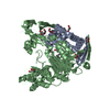
SHEET THE SHEET STRUCTURE OF THIS MOLECULE IS BIFURCATED. IN ORDER TO REPRESENT THIS FEATURE IN ... SHEET THE SHEET STRUCTURE OF THIS MOLECULE IS BIFURCATED. IN ORDER TO REPRESENT THIS FEATURE IN THE SHEET RECORDS BELOW, TWO SHEETS ARE DEFINED.
Mass: 18.015 Da / Num. of mol.: 795 / Source method: isolated from a natural source / Formula: H2O
-
Details
Sequence details
THE AUTHORS INDICATE THAT THE CONFLICT DESCRIBED BELOW IS PROBABLY DUE TO ERROR IN THE SEQUENCE ...THE AUTHORS INDICATE THAT THE CONFLICT DESCRIBED BELOW IS PROBABLY DUE TO ERROR IN THE SEQUENCE DATABASE ANNOTATION, SINCE IN THIS STRUCTURE, NO SIDE CHAIN DENSITY FOR AN ARGININE COULD BE OBSERVED. THE N-TERMINAL SEQUENCING OF THE PROTEIN CRYSTAL CONFIRMS THAT THE SEQUENCE BEGINS AT RESIDUE 61. RESIDUES 479-484 ARE NOT VISIBLE IN THIS CRYSTAL STRUCTURE.
-
Experimental details
-
Experiment
Experiment
Method: X-RAY DIFFRACTION / Number of used crystals: 1
-
Sample preparation
Crystal
Density Matthews: 2.9 Å3/Da / Density % sol: 57.3 %
Resolution: 1.7→87.71 Å / Cor.coef. Fo:Fc: 0.963 / Cor.coef. Fo:Fc free: 0.951 / SU B: 3.529 / SU ML: 0.06 / Cross valid method: THROUGHOUT / ESU R: 0.096 / ESU R Free: 0.094 / Stereochemistry target values: MAXIMUM LIKELIHOOD / Details: HYDROGENS HAVE BEEN ADDED IN THE RIDING POSITIONS.
Rfactor
Num. reflection
% reflection
Selection details
Rfree
0.212
6133
5 %
RANDOM
Rwork
0.185
-
-
-
obs
0.186
116173
98.8 %
-
Solvent computation
Ion probe radii: 0.8 Å / Shrinkage radii: 0.8 Å / VDW probe radii: 1.2 Å
 Movie
Movie Controller

 Yorodumi
 Open data
 Basic information
 Components
 Keywords
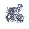
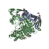
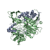
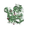
 OXIDOREDUCTASE / FAD /
  Function and homology information
 Authors
 Citation
 Structure visualization
 Downloads & links
 Other downloads


 PDBj
PDBj


 Assembly


 Mass: 96.063 Da / Num. of mol.: 4 / Source method: obtained synthetically / Formula: SO4
 Mass: 22.990 Da / Num. of mol.: 2 / Source method: obtained synthetically / Formula: Na
 Mass: 92.094 Da / Num. of mol.: 7 / Source method: obtained synthetically / Formula: C3H8O3
 Mass: 518.251 Da / Num. of mol.: 2 / Source method: obtained synthetically / Formula: C10H9MoN5O8PS2
 Sample preparation
 / Beamline: BW6 / Wavelength: 1.005
  Processing