 Movie
Movie Controller


+ Open data

- Basic information
| Entry | Database: PDB / ID: 2bhs | ||||||
|---|---|---|---|---|---|---|---|
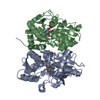
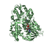
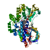
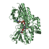
| Title | Crystal Structure of Cysteine Synthase B | ||||||
 Components Components | CYSTEINE SYNTHASE B | ||||||
 Keywords Keywords |  TRANSFERASE / CYSTEINE BIOSYNTHESIS / PLP-DEPENDENT ENZYME / PYRIDOXAL-5'-PHOSPHATE / PYRIDOXAL PHOSPHATE TRANSFERASE / CYSTEINE BIOSYNTHESIS / PLP-DEPENDENT ENZYME / PYRIDOXAL-5'-PHOSPHATE / PYRIDOXAL PHOSPHATE | ||||||
| Function / homology |  Function and homology information Function and homology informationcysteine biosynthetic process via S-sulfo-L-cysteine / L-cysteine catabolic process to pyruvate / cysteine synthase / cysteine biosynthetic process / cysteine synthase activity / L-cysteine desulfhydrase activity / cysteine biosynthetic process from serine / iron-sulfur cluster assembly / pyridoxal phosphate binding / protein homodimerization activity / cytoplasmSimilarity search - Function | ||||||
| Biological species |  ESCHERICHIA COLI (E. coli) ESCHERICHIA COLI (E. coli) | ||||||
| Method | X-RAY DIFFRACTION / SYNCHROTRON / MAD / Resolution: 2.67 Å | ||||||
 Authors Authors | Claus, M.T. / Zocher, G.E. / Maier, T.H.P. / Schulz, G.E. | ||||||
 Citation Citation | Journal: Biochemistry / Year: 2005 Title: Structure of the O-Acetylserine Sulfhydrylase Isoenzyme Cysm from Escherichia Coli Authors: Claus, M.T. / Zocher, G.E. / Maier, T.H.P. / Schulz, G.E. | ||||||
| History |
|
- Structure visualization
| Structure viewer | Molecule: MolmilJmol/JSmol |
|---|
- Downloads & links
-Download
| PDBx/mmCIF format | 2bhs.cif.gz | 224.5 KB | Display | PDBx/mmCIF format |
|---|---|---|---|---|
| PDB format | pdb2bhs.ent.gz | 190 KB | Display | PDB format |
| PDBx/mmJSON format | 2bhs.json.gz | Tree view | PDBx/mmJSON format | |
| Others |  Other downloads Other downloads |
-Validation report
| Arichive directory | https://data.pdbj.org/pub/pdb/validation_reports/bh/2bhsftp://data.pdbj.org/pub/pdb/validation_reports/bh/2bhs | HTTPS FTP |
|---|
-Related structure data


-Links
 PDBj
PDBj
- Assembly
| Deposited unit | 
| ||||||||||||||||
|---|---|---|---|---|---|---|---|---|---|---|---|---|---|---|---|---|---|
| 1 | 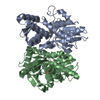
| ||||||||||||||||
| 2 | 
| ||||||||||||||||
| 3 | 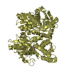
| ||||||||||||||||
| Unit cell |
| ||||||||||||||||
| Noncrystallographic symmetry (NCS) | NCS oper:
|
-Components
| #1: Protein | Mass: 32699.102 Da / Num. of mol.: 4 Source method: isolated from a genetically manipulated source Details: SCHIFF BASE LINK BETWEEN A41 AND PLP320 / Source: (gene. exp.) ESCHERICHIA COLI (E. coli) / Plasmid: PASK-IBA3 / Production host: ESCHERICHIA COLI (E. coli) / Strain (production host): BL21(DE3) / References: UniProt: P16703, cysteine synthase#2: Chemical | ChemComp-PLP / Pyridoxal phosphate  Mass: 247.142 Da / Num. of mol.: 4 / Source method: obtained synthetically / Formula: C8H10NO6P Mass: 247.142 Da / Num. of mol.: 4 / Source method: obtained synthetically / Formula: C8H10NO6P#3: Water | ChemComp-HOH / | Water Mass: 18.015 Da / Num. of mol.: 268 / Source method: isolated from a natural source / Formula: H2O Mass: 18.015 Da / Num. of mol.: 268 / Source method: isolated from a natural source / Formula: H2OCompound details | O(3)-ACETYL-L-SERINE + H(2)S = L-CYSTEINE + ACETATE. | |
|---|
-Experimental details
-Experiment
| Experiment | Method: X-RAY DIFFRACTION / Number of used crystals: 1 |
|---|
- Sample preparation
| Crystal | Density Matthews: 5 Å3/Da / Density % sol: 75 % |
|---|---|
| Crystal grow | pH: 5.6 / Details: pH 5.60 |
-Data collection
| Diffraction | Mean temperature: 100 K |
|---|---|
| Diffraction source | Source: SYNCHROTRON / Site: SLS  / Beamline: X06SA / Wavelength: 0.9795 / Beamline: X06SA / Wavelength: 0.9795 |
| Detector | Type: MARRESEARCH / Detector: CCD |
| Radiation | Protocol: SINGLE WAVELENGTH / Monochromatic (M) / Laue (L): M / Scattering type: x-ray |
| Radiation wavelength | Wavelength: 0.9795 Å / Relative weight: 1 |
| Reflection | Resolution: 2.67→20 Å / Num. obs: 75414 / % possible obs: 99 % / Observed criterion σ(I): 2 / Redundancy: 16.6 % / Rmerge(I) obs: 0.07 / Net I/σ(I): 24.6 |
- Processing
| Software |
| ||||||||||||||||||||||||||||||||||||||||||||||||||||||||||||||||||||||||||||||||||||||||||||||||||||||||||||||||||||||||||||||||||||||||||||||||||||||||||||||||||||||||||||||||||||||
|---|---|---|---|---|---|---|---|---|---|---|---|---|---|---|---|---|---|---|---|---|---|---|---|---|---|---|---|---|---|---|---|---|---|---|---|---|---|---|---|---|---|---|---|---|---|---|---|---|---|---|---|---|---|---|---|---|---|---|---|---|---|---|---|---|---|---|---|---|---|---|---|---|---|---|---|---|---|---|---|---|---|---|---|---|---|---|---|---|---|---|---|---|---|---|---|---|---|---|---|---|---|---|---|---|---|---|---|---|---|---|---|---|---|---|---|---|---|---|---|---|---|---|---|---|---|---|---|---|---|---|---|---|---|---|---|---|---|---|---|---|---|---|---|---|---|---|---|---|---|---|---|---|---|---|---|---|---|---|---|---|---|---|---|---|---|---|---|---|---|---|---|---|---|---|---|---|---|---|---|---|---|---|---|
| Refinement | Method to determine structure: MAD / Resolution: 2.67→19.88 Å / Cor.coef. Fo:Fc: 0.954 / Cor.coef. Fo:Fc free: 0.941 / SU B: 8.318 / SU ML: 0.168 / Cross valid method: THROUGHOUT / ESU R: 0.299 / ESU R Free: 0.241 / Stereochemistry target values: MAXIMUM LIKELIHOOD / Details: HYDROGENS HAVE BEEN ADDED IN THE RIDING POSITIONS.
| ||||||||||||||||||||||||||||||||||||||||||||||||||||||||||||||||||||||||||||||||||||||||||||||||||||||||||||||||||||||||||||||||||||||||||||||||||||||||||||||||||||||||||||||||||||||
| Solvent computation | Ion probe radii: 0.8 Å / Shrinkage radii: 0.8 Å / VDW probe radii: 1.4 Å / Solvent model: BABINET MODEL WITH MASK | ||||||||||||||||||||||||||||||||||||||||||||||||||||||||||||||||||||||||||||||||||||||||||||||||||||||||||||||||||||||||||||||||||||||||||||||||||||||||||||||||||||||||||||||||||||||
| Displacement parameters | Biso mean: 60.3 Å2
| ||||||||||||||||||||||||||||||||||||||||||||||||||||||||||||||||||||||||||||||||||||||||||||||||||||||||||||||||||||||||||||||||||||||||||||||||||||||||||||||||||||||||||||||||||||||
| Refinement step | Cycle: LAST / Resolution: 2.67→19.88 Å
| ||||||||||||||||||||||||||||||||||||||||||||||||||||||||||||||||||||||||||||||||||||||||||||||||||||||||||||||||||||||||||||||||||||||||||||||||||||||||||||||||||||||||||||||||||||||
| Refine LS restraints |
|