 Movie
Movie Controller


+ Open data

- Basic information
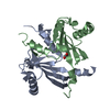
| Entry | Database: PDB / ID: 2bhm | ||||||
|---|---|---|---|---|---|---|---|
| Title | Crystal structure of VirB8 from Brucella suis | ||||||
 Components Components | TYPE IV SECRETION SYSTEM PROTEIN VIRB8 Secretion Secretion | ||||||
 Keywords Keywords | BACTERIAL PROTEIN / BACTERIAL TYPE IV SECRETION | ||||||
| Function / homology |  Function and homology informationprotein secretion by the type IV secretion system / identical protein binding / plasma membrane Function and homology informationprotein secretion by the type IV secretion system / identical protein binding / plasma membraneSimilarity search - Function | ||||||
| Biological species |  BRUCELLA MELITENSIS BIOVAR SUIS (bacteria) BRUCELLA MELITENSIS BIOVAR SUIS (bacteria) | ||||||
| Method | X-RAY DIFFRACTION / SYNCHROTRON / OTHER / Resolution: 2.4 Å | ||||||
 Authors Authors | Bayliss, R. / Baron, C. / Waksman, G. | ||||||
 Citation Citation | Journal: Proc.Natl.Acad.Sci.USA / Year: 2005 Title: Structures of Two Core Subunits of the Bacterial Type Iv Secretion System, Virb8 from Brucella Suis and Comb10 from Helicobacter Pylori Authors: Terradot, L. / Bayliss, R. / Oomen, C. / Leonard, G. / Baron, C. / Waksman, G. | ||||||
| History |
| ||||||
| Remark 650 | HELIX DETERMINATION METHOD: AUTHOR PROVIDED. | ||||||
| Remark 700 | SHEET DETERMINATION METHOD: AUTHOR PROVIDED. |
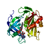
- Structure visualization
| Structure viewer | Molecule: MolmilJmol/JSmol |
|---|
- Downloads & links
-Download
| PDBx/mmCIF format | 2bhm.cif.gz | 140.7 KB | Display | PDBx/mmCIF format |
|---|---|---|---|---|
| PDB format | pdb2bhm.ent.gz | 112.7 KB | Display | PDB format |
| PDBx/mmJSON format | 2bhm.json.gz | Tree view | PDBx/mmJSON format | |
| Others |  Other downloads Other downloads |
-Validation report
| Arichive directory | https://data.pdbj.org/pub/pdb/validation_reports/bh/2bhmftp://data.pdbj.org/pub/pdb/validation_reports/bh/2bhm | HTTPS FTP |
|---|
-Related structure data


-Links
 PDBj
PDBj
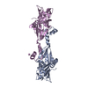
- Assembly
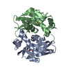
| Deposited unit | 
| ||||||||
|---|---|---|---|---|---|---|---|---|---|
| 1 | 
| ||||||||
| 2 | 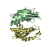
| ||||||||
| 3 | 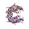
| ||||||||
| Unit cell |
| ||||||||
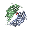
| Details | ACCORDING TO THE AUTHORS OF THIS ENTRY, THE DIMER THAT ISGENERATED BY REMARK 350 BELOW MAY SHOW A LIKELY MODE OFSELF-ASSEMBLY OF VIRB8 WHICH IS KNOWN TO SELF-ASSOCIATETO FORM A LARGE COMPLEX. |
-Components
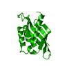
| #1: Protein | Secretion Mass: 18220.145 Da / Num. of mol.: 5 / Fragment: RESIDUES 77-239 Source method: isolated from a genetically manipulated source Source: (gene. exp.) BRUCELLA MELITENSIS BIOVAR SUIS (bacteria)Production host: ESCHERICHIA COLI (E. coli) / Strain (production host): B834 PLYSS / References: UniProt: Q7CEG3#2: Water | ChemComp-HOH / | Water Mass: 18.015 Da / Num. of mol.: 61 / Source method: isolated from a natural source / Formula: H2O Mass: 18.015 Da / Num. of mol.: 61 / Source method: isolated from a natural source / Formula: H2O |
|---|
-Experimental details
-Experiment
| Experiment | Method: X-RAY DIFFRACTION / Number of used crystals: 1 |
|---|
- Sample preparation
| Crystal | Density Matthews: 2.91 Å3/Da / Density % sol: 57.7 % |
|---|
-Data collection
| Diffraction | Mean temperature: 100 K |
|---|---|
| Diffraction source | Source: SYNCHROTRON / Site: ESRF  / Beamline: ID14-4 / Wavelength: 0.9763 / Beamline: ID14-4 / Wavelength: 0.9763 |
| Detector | Type: ADSC CCD / Detector: CCD |
| Radiation | Protocol: SINGLE WAVELENGTH / Monochromatic (M) / Laue (L): M / Scattering type: x-ray |
| Radiation wavelength | Wavelength: 0.9763 Å / Relative weight: 1 |
| Reflection | Resolution: 2.4→22.6 Å / Num. obs: 44402 / % possible obs: 98.8 % / Observed criterion σ(I): 6 / Redundancy: 7.3 % / Rmerge(I) obs: 0.1 / Net I/σ(I): 6.3 |
| Reflection shell | Resolution: 2.4→2.53 Å / Redundancy: 7.2 % / Rmerge(I) obs: 0.44 / Mean I/σ(I) obs: 2.2 / % possible all: 98.8 |
- Processing
| Software |
| ||||||||||||||||||||||||||||||||||||||||||||||||||||||||||||
|---|---|---|---|---|---|---|---|---|---|---|---|---|---|---|---|---|---|---|---|---|---|---|---|---|---|---|---|---|---|---|---|---|---|---|---|---|---|---|---|---|---|---|---|---|---|---|---|---|---|---|---|---|---|---|---|---|---|---|---|---|---|
| Refinement | Method to determine structure: OTHER / Resolution: 2.4→26.6 Å / Data cutoff high absF: 10000 / Cross valid method: THROUGHOUT / σ(F): 0
| ||||||||||||||||||||||||||||||||||||||||||||||||||||||||||||
| Solvent computation | Bsol: 34.2631 Å2 / ksol: 0.357974 e/Å3 | ||||||||||||||||||||||||||||||||||||||||||||||||||||||||||||
| Displacement parameters |
| ||||||||||||||||||||||||||||||||||||||||||||||||||||||||||||
| Refinement step | Cycle: LAST / Resolution: 2.4→26.6 Å
| ||||||||||||||||||||||||||||||||||||||||||||||||||||||||||||
| Refine LS restraints |
| ||||||||||||||||||||||||||||||||||||||||||||||||||||||||||||
| Xplor file |
|
