 Movie
Movie Controller


+ Open data

- Basic information
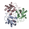
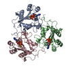
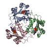
| Entry | Database: PDB / ID: 2bef | ||||||
|---|---|---|---|---|---|---|---|
| Title | CRYSTAL STRUCTURE OF NDP KINASE COMPLEXED WITH MG, ADP, AND BEF3 | ||||||
 Components Components | NUCLEOSIDE DIPHOSPHATE KINASE Nucleoside-diphosphate kinase Nucleoside-diphosphate kinase | ||||||
 Keywords Keywords | PHOSPHOTRANSFERASE / NUCLEOSIDE DIPHOSPHATE KINASE / PHOSPHORYL TRANSFER / BERYLLIUM FLUORIDE | ||||||
| Function / homology |  Function and homology information Function and homology informationdGTP biosynthetic process from dGDP / Azathioprine ADME / Ribavirin ADME / asexual reproduction / Interconversion of nucleotide di- and triphosphates / Neutrophil degranulation / nucleoside triphosphate biosynthetic process / negative regulation of pinocytosis / nucleoside-diphosphate kinase / UTP biosynthetic process ...dGTP biosynthetic process from dGDP / Azathioprine ADME / Ribavirin ADME / asexual reproduction / Interconversion of nucleotide di- and triphosphates / Neutrophil degranulation / nucleoside triphosphate biosynthetic process / negative regulation of pinocytosis / nucleoside-diphosphate kinase / UTP biosynthetic process / CTP biosynthetic process / negative regulation of exocytosis / negative regulation of phagocytosis / GTP biosynthetic process / nucleoside diphosphate kinase activity / translational elongation / phagocytic vesicle / secretory granule / response to bacterium / actin cytoskeleton organization / cytoskeleton / ribosome / G protein-coupled receptor signaling pathway / phosphorylation / ATP binding / metal ion binding / plasma membrane / cytoplasmSimilarity search - Function | ||||||
| Biological species |  Dictyostelium discoideum (eukaryote) Dictyostelium discoideum (eukaryote) | ||||||
| Method | X-RAY DIFFRACTION / MOLECULAR REPLACEMENT / Resolution: 2.3 Å | ||||||
 Authors Authors | Xu, Y.W. / Cherfils, J. | ||||||
 Citation Citation | Journal: Proc.Natl.Acad.Sci.USA / Year: 1997 Title: AlF3 mimics the transition state of protein phosphorylation in the crystal structure of nucleoside diphosphate kinase and MgADP. Authors: Xu, Y.W. / Morera, S. / Janin, J. / Cherfils, J. | ||||||
| History |
|
- Structure visualization
| Structure viewer | Molecule: MolmilJmol/JSmol |
|---|
- Downloads & links
-Download
| PDBx/mmCIF format | 2bef.cif.gz | 100.5 KB | Display | PDBx/mmCIF format |
|---|---|---|---|---|
| PDB format | pdb2bef.ent.gz | 83 KB | Display | PDB format |
| PDBx/mmJSON format | 2bef.json.gz | Tree view | PDBx/mmJSON format | |
| Others |  Other downloads Other downloads |
-Validation report
| Arichive directory | https://data.pdbj.org/pub/pdb/validation_reports/be/2befftp://data.pdbj.org/pub/pdb/validation_reports/be/2bef | HTTPS FTP |
|---|
-Related structure data

-Links
 PDBj
PDBj


- Assembly
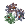
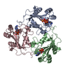
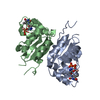
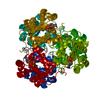
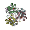
| Deposited unit | 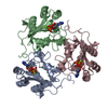
| ||||||||
|---|---|---|---|---|---|---|---|---|---|
| 1 | 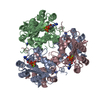
| ||||||||
| Unit cell |
| ||||||||
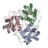
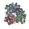
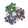
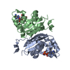
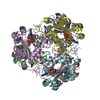
| Details | THE DICTYOSTELIUM NDP KINASE HEXAMER IS RECONSTITUTED FROM THE TRIMER IN THE ASYMMETRIC UNIT FROM CRYSTALLOGRAPHIC SYMMETRIES. THE POSITION OF BE++ WAS MODELLED IN TETRAHEDRAL CONFORMATION FROM THE ELECTRON DENSITY OF THE 3 FLUORINE ATOMS. |
-Components
| #1: Protein | Nucleoside-diphosphate kinase Mass: 16816.336 Da / Num. of mol.: 3 Source method: isolated from a genetically manipulated source Details: ADP, BEF3, MG++ / Source: (gene. exp.) Dictyostelium discoideum (eukaryote) / Production host:  Escherichia coli (E. coli) / References: UniProt: P22887, nucleoside-diphosphate kinase Escherichia coli (E. coli) / References: UniProt: P22887, nucleoside-diphosphate kinase#2: Chemical |   Mass: 24.305 Da / Num. of mol.: 3 / Source method: obtained synthetically / Formula: Mg Mass: 24.305 Da / Num. of mol.: 3 / Source method: obtained synthetically / Formula: Mg#3: Chemical |   Mass: 66.007 Da / Num. of mol.: 3 / Source method: obtained synthetically / Formula: BeF3 Mass: 66.007 Da / Num. of mol.: 3 / Source method: obtained synthetically / Formula: BeF3#4: Chemical | Adenosine diphosphate  Mass: 427.201 Da / Num. of mol.: 3 / Source method: obtained synthetically / Formula: C10H15N5O10P2 / Comment: ADP, energy-carrying molecule*YM Mass: 427.201 Da / Num. of mol.: 3 / Source method: obtained synthetically / Formula: C10H15N5O10P2 / Comment: ADP, energy-carrying molecule*YM#5: Water | ChemComp-HOH / | Water Mass: 18.015 Da / Num. of mol.: 220 / Source method: isolated from a natural source / Formula: H2O Mass: 18.015 Da / Num. of mol.: 220 / Source method: isolated from a natural source / Formula: H2O |
|---|
-Experimental details
-Experiment
| Experiment | Method: X-RAY DIFFRACTION / Number of used crystals: 1 |
|---|
- Sample preparation
| Crystal | Density Matthews: 2.25 Å3/Da / Density % sol: 38 % / Description: 100% IDENTITY | ||||||||||||||||||||||||||||||||||||||||||||||||
|---|---|---|---|---|---|---|---|---|---|---|---|---|---|---|---|---|---|---|---|---|---|---|---|---|---|---|---|---|---|---|---|---|---|---|---|---|---|---|---|---|---|---|---|---|---|---|---|---|---|
| Crystal grow | pH: 7.5 Details: HANGING DROP, SEEDING, 32% PEG550, 50MM TRIS PH 7.5, 20MM MGCL2, 25MM NAF, 1MM BECL2 | ||||||||||||||||||||||||||||||||||||||||||||||||
| Crystal | *PLUS | ||||||||||||||||||||||||||||||||||||||||||||||||
| Crystal grow | *PLUS Temperature: 20 ℃ / pH: 7.4 / Method: vapor diffusion, hanging drop | ||||||||||||||||||||||||||||||||||||||||||||||||
| Components of the solutions | *PLUS
|
-Data collection
| Diffraction | Mean temperature: 277 K |
|---|---|
| Diffraction source | Source: ROTATING ANODE / Type: RIGAKU / Wavelength: 1.5418 |
| Detector | Type: RIGAKU RAXIS / Detector: IMAGE PLATE / Date: Apr 1, 1996 |
| Radiation | Monochromatic (M) / Laue (L): M / Scattering type: x-ray |
| Radiation wavelength | Wavelength: 1.5418 Å / Relative weight: 1 |
| Reflection | Resolution: 2.3→15 Å / Num. obs: 20554 / % possible obs: 98 % / Observed criterion σ(I): 2 / Redundancy: 2.5 % / Biso Wilson estimate: 33 Å2 / Rsym value: 0.053 / Net I/σ(I): 13.1 |
| Reflection shell | Resolution: 2.3→2.36 Å / Redundancy: 2.2 % / Rmerge(I) obs: 0.207 / Mean I/σ(I) obs: 3.6 / % possible all: 94 |
| Reflection | *PLUS Num. measured all: 51271 / Rmerge(I) obs: 0.053 |
- Processing
| Software |
| ||||||||||||||||||||||||||||||||||||||||||||||||||||||||||||
|---|---|---|---|---|---|---|---|---|---|---|---|---|---|---|---|---|---|---|---|---|---|---|---|---|---|---|---|---|---|---|---|---|---|---|---|---|---|---|---|---|---|---|---|---|---|---|---|---|---|---|---|---|---|---|---|---|---|---|---|---|---|
| Refinement | Method to determine structure: MOLECULAR REPLACEMENT Starting model: NDPK + MGADP + ALF3, WITH MGADP AND ALF3 REMOVED FROM THE MODEL Resolution: 2.3→15 Å / σ(F): 2
| ||||||||||||||||||||||||||||||||||||||||||||||||||||||||||||
| Displacement parameters | Biso mean: 18.6 Å2 | ||||||||||||||||||||||||||||||||||||||||||||||||||||||||||||
| Refine analyze | Luzzati coordinate error obs: 0.35 Å / Luzzati d res low obs: 4 Å / Luzzati sigma a obs: 0.3 Å | ||||||||||||||||||||||||||||||||||||||||||||||||||||||||||||
| Refinement step | Cycle: LAST / Resolution: 2.3→15 Å
| ||||||||||||||||||||||||||||||||||||||||||||||||||||||||||||
| Refine LS restraints |
| ||||||||||||||||||||||||||||||||||||||||||||||||||||||||||||
| Software | *PLUS Name: X-PLOR / Version: 3.8 / Classification: refinement | ||||||||||||||||||||||||||||||||||||||||||||||||||||||||||||
| Refinement | *PLUS Num. reflection obs: 20058 | ||||||||||||||||||||||||||||||||||||||||||||||||||||||||||||
| Solvent computation | *PLUS | ||||||||||||||||||||||||||||||||||||||||||||||||||||||||||||
| Displacement parameters | *PLUS | ||||||||||||||||||||||||||||||||||||||||||||||||||||||||||||
| Refine LS restraints | *PLUS
|
