| Entry | Database: PDB / ID: 2b98
|
|---|
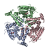
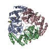
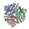
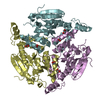
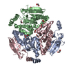
| Title | Crystal Structure of an archaeal pentameric riboflavin synthase |
|---|
 Components Components | Riboflavin synthase |
|---|
 Keywords Keywords | TRANSFERASE / lumazine riboflavin |
|---|
| Function / homology |  Function and homology information Function and homology information |
|---|
| Biological species |   Methanocaldococcus jannaschii (archaea) Methanocaldococcus jannaschii (archaea) |
|---|
| Method | X-RAY DIFFRACTION / MOLECULAR REPLACEMENT / Resolution: 2.3 Å |
|---|
 Authors Authors | Ramsperger, A. / Augustin, M. / Schott, A.K. / Gerhardt, S. / Krojer, T. / Eisenreich, W. / Illarionov, B. / Cushman, M. / Bacher, A. / Huber, R. / Fischer, M. |
|---|
 Citation Citation | Journal: J.Biol.Chem. / Year: 2006
Title: Crystal Structure of an Archaeal Pentameric Riboflavin Synthase in Complex with a Substrate Analog Inhibitor: stereochemical implications
Authors: Ramsperger, A. / Augustin, M. / Schott, A.K. / Gerhardt, S. / Krojer, T. / Eisenreich, W. / Illarionov, B. / Cushman, M. / Bacher, A. / Huber, R. / Fischer, M. |
|---|
| History | | Deposition | Oct 11, 2005 | Deposition site: RCSB / Processing site: RCSB |
|---|
| Revision 1.0 | Nov 8, 2005 | Provider: repository / Type: Initial release |
|---|
| Revision 1.1 | May 1, 2008 | Group: Version format compliance |
|---|
| Revision 1.2 | Jul 13, 2011 | Group: Advisory / Version format compliance |
|---|
| Revision 1.3 | Feb 14, 2024 | Group: Data collection / Database references / Refinement description
Category: chem_comp_atom / chem_comp_bond ...chem_comp_atom / chem_comp_bond / database_2 / struct_ncs_dom_lim
Item: _database_2.pdbx_DOI / _database_2.pdbx_database_accession ..._database_2.pdbx_DOI / _database_2.pdbx_database_accession / _struct_ncs_dom_lim.beg_auth_comp_id / _struct_ncs_dom_lim.end_auth_comp_id |
|---|
|
|---|
 Movie
Movie Controller


 Open data
 Basic information
 Structure visualization
 Downloads & links
 Other downloads


 PDBj
PDBj Assembly