 Movie
Movie Controller


+ Open data

- Basic information
| Entry | Database: PDB / ID: 2b34 | ||||||
|---|---|---|---|---|---|---|---|
| Title | Structure of MAR1 Ribonuclease from Caenorhabditis elegans | ||||||
 Components Components | MAR1 Ribonuclease | ||||||
 Keywords Keywords |  HYDROLASE / Isochorismatase Family / Structural Genomics / PSI / Protein Structure Initiative / Southeast Collaboratory for Structural Genomics / SECSG HYDROLASE / Isochorismatase Family / Structural Genomics / PSI / Protein Structure Initiative / Southeast Collaboratory for Structural Genomics / SECSG | ||||||
| Function / homology | Isochorismatase-like / Isochorismatase-like / Isochorismatase-like superfamily / Isochorismatase family / Rossmann fold / 3-Layer(aba) Sandwich / cytoplasm / Alpha Beta / Isochorismatase domain-containing protein Function and homology information Function and homology information | ||||||
| Biological species |  Caenorhabditis elegans (invertebrata) Caenorhabditis elegans (invertebrata) | ||||||
| Method | X-RAY DIFFRACTION / MOLECULAR REPLACEMENT / Resolution: 2.141 Å | ||||||
 Authors Authors | Schormann, N. / Karpova, E. / Li, S. / Symersky, J. / Zhang, Y. / Lu, S. / Zhou, Q. / Lin, G. / Cao, Z. / Luo, M. ...Schormann, N. / Karpova, E. / Li, S. / Symersky, J. / Zhang, Y. / Lu, S. / Zhou, Q. / Lin, G. / Cao, Z. / Luo, M. / Qiu, S. / Luan, C.-H. / Luo, D. / Huang, W. / Shang, Q. / McKinstry, A. / An, J. / Tsao, J. / Carson, M. / Stinnett, M. / Chen, Y. / Johnson, D. / Gary, R. / Arabshahi, A. / Bunzel, R. / Bray, T. / DeLucas, L. / Southeast Collaboratory for Structural Genomics (SECSG) | ||||||
 Citation Citation | Journal: To be Published Title: Structure of MAR1 Ribonuclease from Caenorhabditis elegans Authors: Schormann, N. / Karpova, E. / Li, S. / Symersky, J. / Zhang, Y. / Lu, S. / Zhou, Q. / Lin, G. / Cao, Z. / Luo, M. / Qiu, S. / Luan, C.-H. / Luo, D. / Huang, W. / Shang, Q. / McKinstry, A. / ...Authors: Schormann, N. / Karpova, E. / Li, S. / Symersky, J. / Zhang, Y. / Lu, S. / Zhou, Q. / Lin, G. / Cao, Z. / Luo, M. / Qiu, S. / Luan, C.-H. / Luo, D. / Huang, W. / Shang, Q. / McKinstry, A. / An, J. / Tsao, J. / Carson, M. / Stinnett, M. / Chen, Y. / Johnson, D. / Gary, R. / Arabshahi, A. / Bunzel, R. / Bray, T. / DeLucas, L. | ||||||
| History |
|
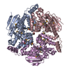
- Structure visualization
| Structure viewer | Molecule: MolmilJmol/JSmol |
|---|
- Downloads & links
-Download
| PDBx/mmCIF format | 2b34.cif.gz | 302.8 KB | Display | PDBx/mmCIF format |
|---|---|---|---|---|
| PDB format | pdb2b34.ent.gz | 248.6 KB | Display | PDB format |
| PDBx/mmJSON format | 2b34.json.gz | Tree view | PDBx/mmJSON format | |
| Others |  Other downloads Other downloads |
-Validation report
| Arichive directory | https://data.pdbj.org/pub/pdb/validation_reports/b3/2b34ftp://data.pdbj.org/pub/pdb/validation_reports/b3/2b34 | HTTPS FTP |
|---|
-Related structure data
| Related structure data |  1x9gS S: Starting model for refinement |
|---|---|
| Similar structure data | |
| Other databases |


-Links
 PDBj
PDBj- Assembly
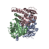
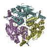
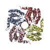
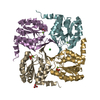
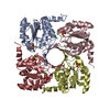
| Deposited unit | 
| ||||||||
|---|---|---|---|---|---|---|---|---|---|
| 1 | 
| ||||||||
| 2 | 
| ||||||||
| 3 | 
| ||||||||
| Unit cell |
| ||||||||
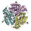
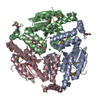
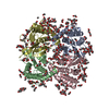
| Details | The asymmetric unit contains two tetramers in an octameric assembly, with the second tetramer off-center of the first. The biological unit is probably the tetramer. |
-Components
| #1: Protein | Mass: 21877.426 Da / Num. of mol.: 8 Source method: isolated from a genetically manipulated source Source: (gene. exp.) Caenorhabditis elegans (invertebrata) / Strain: Bristol N2 / Plasmid: pET28b / Production host:  Escherichia coli (E. coli) / Strain (production host): BL21 AI / References: UniProt: Q20062 Escherichia coli (E. coli) / Strain (production host): BL21 AI / References: UniProt: Q20062#2: Water | ChemComp-HOH / | Water Mass: 18.015 Da / Num. of mol.: 607 / Source method: isolated from a natural source / Formula: H2O Mass: 18.015 Da / Num. of mol.: 607 / Source method: isolated from a natural source / Formula: H2O |
|---|
-Experimental details
-Experiment
| Experiment | Method: X-RAY DIFFRACTION / Number of used crystals: 1 |
|---|
- Sample preparation
| Crystal | Density Matthews: 2.2 Å3/Da / Density % sol: 44.2 % |
|---|---|
| Crystal grow | Temperature: 291 K / Method: vapor diffusion, hanging drop / pH: 8 Details: 20% PEG3350, 0.2M sodium acetate, 0.1M Tris , pH 8.0, VAPOR DIFFUSION, HANGING DROP, temperature 291K |
-Data collection
| Diffraction | Mean temperature: 103 K |
|---|---|
| Diffraction source | Source: ROTATING ANODE / Type: RIGAKU RU300 / Wavelength: 1.5418 Å |
| Detector | Type: RIGAKU RAXIS IV / Detector: IMAGE PLATE / Date: Mar 1, 2005 / Details: Mirrors |
| Radiation | Monochromator: Graphite / Protocol: SINGLE WAVELENGTH / Monochromatic (M) / Laue (L): M / Scattering type: x-ray |
| Radiation wavelength | Wavelength: 1.5418 Å / Relative weight: 1 |
| Reflection | Resolution: 2.14→50 Å / Num. all: 84631 / Num. obs: 84631 / % possible obs: 98.3 % / Observed criterion σ(I): 1 / Redundancy: 3.5 % / Biso Wilson estimate: 18.7 Å2 / Rmerge(I) obs: 0.062 / Net I/σ(I): 23.4 |
| Reflection shell | Resolution: 2.14→2.22 Å / Redundancy: 3.3 % / Rmerge(I) obs: 0.168 / Num. unique all: 8022 / % possible all: 94.7 |
- Processing
| Software |
| ||||||||||||||||||||||||||||||||||||||||||||||||||||||||||||||||||||||||||||||||||||||||||||||||||||||||||||||||||||||||||||||||||||||||||||||||||||||||||||||||||||||||||
|---|---|---|---|---|---|---|---|---|---|---|---|---|---|---|---|---|---|---|---|---|---|---|---|---|---|---|---|---|---|---|---|---|---|---|---|---|---|---|---|---|---|---|---|---|---|---|---|---|---|---|---|---|---|---|---|---|---|---|---|---|---|---|---|---|---|---|---|---|---|---|---|---|---|---|---|---|---|---|---|---|---|---|---|---|---|---|---|---|---|---|---|---|---|---|---|---|---|---|---|---|---|---|---|---|---|---|---|---|---|---|---|---|---|---|---|---|---|---|---|---|---|---|---|---|---|---|---|---|---|---|---|---|---|---|---|---|---|---|---|---|---|---|---|---|---|---|---|---|---|---|---|---|---|---|---|---|---|---|---|---|---|---|---|---|---|---|---|---|---|---|---|
| Refinement | Method to determine structure: MOLECULAR REPLACEMENT Starting model: The starting model was created by the Swiss-Model Server based on PDB entry 1X9G using the C. elegans target sequence (F35G2.2) Resolution: 2.141→41.67 Å / Cor.coef. Fo:Fc: 0.923 / Cor.coef. Fo:Fc free: 0.896 / SU B: 5.843 / SU ML: 0.157 / Isotropic thermal model: Isotropic / Cross valid method: THROUGHOUT / σ(F): 0 / ESU R: 0.32 / ESU R Free: 0.23 / Stereochemistry target values: MAXIMUM LIKELIHOOD / Details: HYDROGENS HAVE BEEN ADDED IN THE RIDING POSITIONS
| ||||||||||||||||||||||||||||||||||||||||||||||||||||||||||||||||||||||||||||||||||||||||||||||||||||||||||||||||||||||||||||||||||||||||||||||||||||||||||||||||||||||||||
| Solvent computation | Ion probe radii: 0.8 Å / Shrinkage radii: 0.8 Å / VDW probe radii: 1.2 Å / Solvent model: MASK | ||||||||||||||||||||||||||||||||||||||||||||||||||||||||||||||||||||||||||||||||||||||||||||||||||||||||||||||||||||||||||||||||||||||||||||||||||||||||||||||||||||||||||
| Displacement parameters | Biso mean: 25.126 Å2
| ||||||||||||||||||||||||||||||||||||||||||||||||||||||||||||||||||||||||||||||||||||||||||||||||||||||||||||||||||||||||||||||||||||||||||||||||||||||||||||||||||||||||||
| Refine analyze |
| ||||||||||||||||||||||||||||||||||||||||||||||||||||||||||||||||||||||||||||||||||||||||||||||||||||||||||||||||||||||||||||||||||||||||||||||||||||||||||||||||||||||||||
| Refinement step | Cycle: LAST / Resolution: 2.141→41.67 Å
| ||||||||||||||||||||||||||||||||||||||||||||||||||||||||||||||||||||||||||||||||||||||||||||||||||||||||||||||||||||||||||||||||||||||||||||||||||||||||||||||||||||||||||
| Refine LS restraints |
| ||||||||||||||||||||||||||||||||||||||||||||||||||||||||||||||||||||||||||||||||||||||||||||||||||||||||||||||||||||||||||||||||||||||||||||||||||||||||||||||||||||||||||
| LS refinement shell | Resolution: 2.141→2.196 Å / Rfactor Rfree error: 0.01 / Total num. of bins used: 20
|
