 Movie
Movie Controller

[English] 日本語
 Yorodumi
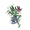
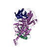
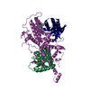
- PDB-2ayu: The structure of nucleosome assembly protein suggests a mechanism... -
+ Open data

- Basic information
| Entry | Database: PDB / ID: 2ayu | ||||||
|---|---|---|---|---|---|---|---|
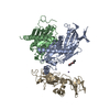
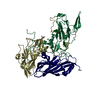
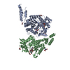
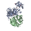
| Title | The structure of nucleosome assembly protein suggests a mechanism for histone binding and shuttling | ||||||
 Components Components | Nucleosome assembly protein | ||||||
 Keywords Keywords |  CHAPERONE / Nucleosome assembly protein 1 (NAP1) / Histone chaperone CHAPERONE / Nucleosome assembly protein 1 (NAP1) / Histone chaperone | ||||||
| Function / homology |  Function and homology information Function and homology informationprotein localization to cell division site after cytokinesis / Gin4 complex / cellular bud neck septin collar / budding cell bud growth / : / septin ring assembly / septum digestion after cytokinesis / nucleosome disassembly / NLS-bearing protein import into nucleus / cell division site ...protein localization to cell division site after cytokinesis / Gin4 complex / cellular bud neck septin collar / budding cell bud growth / : / septin ring assembly / septum digestion after cytokinesis / nucleosome disassembly / NLS-bearing protein import into nucleus / cell division site / enzyme activator activity / positive regulation of microtubule polymerization / cyclin binding / positive regulation of transcription elongation by RNA polymerase II / ribosomal small subunit biogenesis / nucleosome assembly / unfolded protein binding / histone binding / chromatin binding / chromatin / DNA binding / identical protein binding / nucleus / cytoplasmSimilarity search - Function | ||||||
| Biological species |  Saccharomyces cerevisiae (brewer's yeast) Saccharomyces cerevisiae (brewer's yeast) | ||||||
| Method | X-RAY DIFFRACTION / SYNCHROTRON / MAD / Resolution: 3 Å | ||||||
 Authors Authors | Park, Y.J. / Luger, K. | ||||||
 Citation Citation | Journal: Proc.Natl.Acad.Sci.Usa / Year: 2006 Title: The structure of nucleosome assembly protein 1. Authors: Park, Y.J. / Luger, K. | ||||||
| History |
|
- Structure visualization
| Structure viewer | Molecule: MolmilJmol/JSmol |
|---|
- Downloads & links
-Download
| PDBx/mmCIF format | 2ayu.cif.gz | 60.2 KB | Display | PDBx/mmCIF format |
|---|---|---|---|---|
| PDB format | pdb2ayu.ent.gz | 46.8 KB | Display | PDB format |
| PDBx/mmJSON format | 2ayu.json.gz | Tree view | PDBx/mmJSON format | |
| Others |  Other downloads Other downloads |
-Validation report
| Arichive directory | https://data.pdbj.org/pub/pdb/validation_reports/ay/2ayuftp://data.pdbj.org/pub/pdb/validation_reports/ay/2ayu | HTTPS FTP |
|---|
-Related structure data
| Similar structure data |
|---|


-Links
 PDBj
PDBj
- Assembly
| Deposited unit | 
| ||||||||
|---|---|---|---|---|---|---|---|---|---|
| 1 | 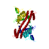
| ||||||||
| Unit cell |
|
-Components
| #1: Protein | Mass: 47930.383 Da / Num. of mol.: 1 Source method: isolated from a genetically manipulated source Source: (gene. exp.) Saccharomyces cerevisiae (brewer's yeast)Gene: NAP1 / Species (production host): Escherichia coli / Production host:  Escherichia coli BL21(DE3) (bacteria) / Strain (production host): BL21DE3 / References: UniProt: P25293 Escherichia coli BL21(DE3) (bacteria) / Strain (production host): BL21DE3 / References: UniProt: P25293 |
|---|
-Experimental details
-Experiment
| Experiment | Method: X-RAY DIFFRACTION / Number of used crystals: 1 |
|---|
- Sample preparation
| Crystal | Density Matthews: 3.45 Å3/Da / Density % sol: 64.35 % |
|---|---|
| Crystal grow | Temperature: 292 K / Method: vapor diffusion, sitting drop / pH: 4.5 Details: mono-ammonium dihydrogen phosphate, pH 4.5, VAPOR DIFFUSION, SITTING DROP, temperature 292K |
-Data collection
| Diffraction |
| |||||||||||||||
|---|---|---|---|---|---|---|---|---|---|---|---|---|---|---|---|---|
| Diffraction source |
| |||||||||||||||
| Detector |
| |||||||||||||||
| Radiation |
| |||||||||||||||
| Radiation wavelength |
| |||||||||||||||
| Reflection | Resolution: 3→50 Å |

- Processing
| Software |
| |||||||||||||||
|---|---|---|---|---|---|---|---|---|---|---|---|---|---|---|---|---|
| Refinement | Method to determine structure: MAD / Resolution: 3→50 Å / Rfactor Rfree: 0.28 / Rfactor Rwork: 0.25 / σ(F): 0 / σ(I): 0 | |||||||||||||||
| Refinement step | Cycle: LAST / Resolution: 3→50 Å
| |||||||||||||||
| Refine LS restraints |
|
