- PDB-2awa: Crystal structure of DNA polymerase III, beta chain (EC 2.7.7.7) ... -
+
Open data
ID or keywords:
Loading...
-
Basic information
Entry
Database: PDB / ID: 2awa
Title
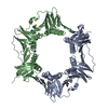
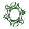
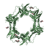
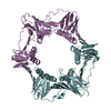
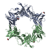
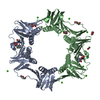
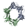
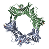
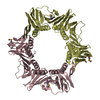
Crystal structure of DNA polymerase III, beta chain (EC 2.7.7.7) (np_344555.1) from STREPTOCOCCUS PNEUMONIAE TIGR4 at 2.50 A resolution
Components
DNA polymerase III, beta chain
Keywords
TRANSFERASE / np_344555.1 / DNA polymerase III / beta chain (EC 2.7.7.7) / Structural Genomics / Joint Center for Structural Genomics / JCSG / Protein Structure Initiative / PSI
Function / homology
Function and homology information
DNA polymerase III complex / 3'-5' exonuclease activity / DNA replication / DNA-directed DNA polymerase activity / DNA binding / cytoplasm Similarity search - Function
DNA Polymerase III; Chain A, domain 2 / DNA Polymerase III, subunit A, domain 2 / Box / Proliferating Cell Nuclear Antigen / Proliferating Cell Nuclear Antigen - #10 / DNA polymerase III, beta sliding clamp / DNA polymerase III, beta sliding clamp, N-terminal / DNA polymerase III, beta sliding clamp, C-terminal / DNA polymerase III, beta sliding clamp, central / DNA polymerase III beta subunit, N-terminal domain ...DNA Polymerase III; Chain A, domain 2 / DNA Polymerase III, subunit A, domain 2 / Box / Proliferating Cell Nuclear Antigen / Proliferating Cell Nuclear Antigen - #10 / DNA polymerase III, beta sliding clamp / DNA polymerase III, beta sliding clamp, N-terminal / DNA polymerase III, beta sliding clamp, C-terminal / DNA polymerase III, beta sliding clamp, central / DNA polymerase III beta subunit, N-terminal domain / DNA polymerase III beta subunit, central domain / DNA polymerase III beta subunit, C-terminal domain / DNA polymerase III beta subunit / Roll / Alpha Beta Similarity search - Domain/homology
A: DNA polymerase III, beta chain B: DNA polymerase III, beta chain C: DNA polymerase III, beta chain D: DNA polymerase III, beta chain hetero molecules
Mass: 18.015 Da / Num. of mol.: 251 / Source method: isolated from a natural source / Formula: H2O
-
Experimental details
-
Experiment
Experiment
Method: X-RAY DIFFRACTION / Number of used crystals: 1
-
Sample preparation
Crystal
Density Matthews: 2.76 Å3/Da / Density % sol: 55.16 % Description: THE EXPERIMENTAL MAD MAP IS POOR WITHOUT NCS AVERAGING. A MONOMER POLY-ALANINE MODEL OF 1MMI IS USED AS TEMPLATE FOR THE LOCATION OF FOUR MONOMERS IN THE EXPERIMENTAL MAP. THE MAD MAP IS ...Description: THE EXPERIMENTAL MAD MAP IS POOR WITHOUT NCS AVERAGING. A MONOMER POLY-ALANINE MODEL OF 1MMI IS USED AS TEMPLATE FOR THE LOCATION OF FOUR MONOMERS IN THE EXPERIMENTAL MAP. THE MAD MAP IS SIGNIFICANTLY IMPROVED WITH NCS AVERAGING WHICH ALLOWS THE COMPLETION OF THE MODEL FROM THE INITIAL PHASED MOLECULAR REPLACEMENT SOLUTION. THE IMPROVED PHASES RESULTING FROM NCS AVERAGING WERE USED AS PHASE RESTRAINTS IN THE REFMAC REFINEMENT.
Resolution: 2.5→30 Å / Cor.coef. Fo:Fc: 0.955 / Cor.coef. Fo:Fc free: 0.924 / SU B: 21.693 / SU ML: 0.229 / TLS residual ADP flag: LIKELY RESIDUAL / Cross valid method: THROUGHOUT / ESU R: 0.535 / ESU R Free: 0.292 Stereochemistry target values: MAXIMUM LIKELIHOOD WITH PHASES Details: 1. HYDROGENS HAVE BEEN ADDED IN THE RIDING POSITIONS 2. MANY SURFACE SIDE CHAINS ARE TRIMMED DUE TO LACK OF DEFINITIVE ELECTRON DENSITIES.
Rfactor
Num. reflection
% reflection
Selection details
Rfree
0.25
3044
5.1 %
RANDOM
Rwork
0.192
-
-
-
all
0.195
-
-
-
obs
-
56494
48.91 %
-
Solvent computation
Ion probe radii: 0.8 Å / Shrinkage radii: 0.8 Å / VDW probe radii: 1.2 Å / Solvent model: BABINET MODEL WITH MASK
In the structure databanks used in Yorodumi, some data are registered as the other names, "COVID-19 virus" and "2019-nCoV". Here are the details of the virus and the list of structure data.
Jan 31, 2019. EMDB accession codes are about to change! (news from PDBe EMDB page)
EMDB accession codes are about to change! (news from PDBe EMDB page)
The allocation of 4 digits for EMDB accession codes will soon come to an end. Whilst these codes will remain in use, new EMDB accession codes will include an additional digit and will expand incrementally as the available range of codes is exhausted. The current 4-digit format prefixed with “EMD-” (i.e. EMD-XXXX) will advance to a 5-digit format (i.e. EMD-XXXXX), and so on. It is currently estimated that the 4-digit codes will be depleted around Spring 2019, at which point the 5-digit format will come into force.
The EM Navigator/Yorodumi systems omit the EMD- prefix.
Related info.:Q: What is EMD? / ID/Accession-code notation in Yorodumi/EM Navigator
Yorodumi is a browser for structure data from EMDB, PDB, SASBDB, etc.
This page is also the successor to EM Navigator detail page, and also detail information page/front-end page for Omokage search.
The word "yorodu" (or yorozu) is an old Japanese word meaning "ten thousand". "mi" (miru) is to see.
Related info.:EMDB / PDB / SASBDB / Comparison of 3 databanks / Yorodumi Search / Aug 31, 2016. New EM Navigator & Yorodumi / Yorodumi Papers / Jmol/JSmol / Function and homology information / Changes in new EM Navigator and Yorodumi
 Movie
Movie Controller

 Yorodumi
 Open data
 Basic information
 Components
 Keywords
 Function and homology information

 Authors
 Citation
 Structure visualization
 Downloads & links
 Other downloads
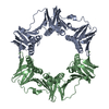


 PDBj
PDBj
 Assembly

 Mass: 92.094 Da / Num. of mol.: 1 / Source method: obtained synthetically / Formula: C3H8O3
 Mass: 18.015 Da / Num. of mol.: 251 / Source method: isolated from a natural source / Formula: H2O
 Sample preparation
 / Beamline: BL9-2 / Wavelength: 0.97934, 0.91837, 0.97923
 Processing