 Movie
Movie Controller

[English] 日本語
 Yorodumi
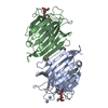
- PDB-2arx: Pterocarpus angolensis seed lectin in complex with the decasaccha... -
+ Open data

- Basic information
| Entry | Database: PDB / ID: 2arx | |||||||||
|---|---|---|---|---|---|---|---|---|---|---|
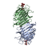
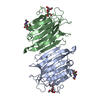
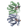
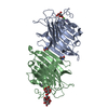
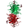
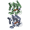
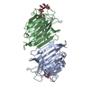
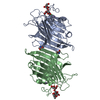
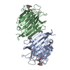
| Title | Pterocarpus angolensis seed lectin in complex with the decasaccharide NA2F | |||||||||
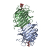
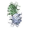
 Components Components | lectin | |||||||||
 Keywords Keywords | SUGAR BINDING PROTEIN / lectin / carbohydrate / legume lectin | |||||||||
| Function / homology |  Function and homology information Function and homology information | |||||||||
| Biological species |  Pterocarpus angolensis (plant) Pterocarpus angolensis (plant) | |||||||||
| Method | X-RAY DIFFRACTION / SYNCHROTRON / MOLECULAR REPLACEMENT / Resolution: 2 Å | |||||||||
 Authors Authors | Buts, L. / Garcia-Pino, A. / Imberty, A. / Amiot, N. / Boons, G.-J. / Lah, J. / Versees, W. / Wyns, L. / Loris, R. | |||||||||
 Citation Citation | Journal: Febs J. / Year: 2006 Title: Structural basis for the recognition of complex-type biantennary oligosaccharides by Pterocarpus angolensis lectin. Authors: Buts, L. / Garcia-Pino, A. / Imberty, A. / Amiot, N. / Boons, G.J. / Beeckmans, S. / Versees, W. / Wyns, L. / Loris, R. | |||||||||
| History |
|
- Structure visualization
| Structure viewer | Molecule: MolmilJmol/JSmol |
|---|
- Downloads & links
-Download
| PDBx/mmCIF format | 2arx.cif.gz | 115.6 KB | Display | PDBx/mmCIF format |
|---|---|---|---|---|
| PDB format | pdb2arx.ent.gz | 86.3 KB | Display | PDB format |
| PDBx/mmJSON format | 2arx.json.gz | Tree view | PDBx/mmJSON format | |
| Others |  Other downloads Other downloads |
-Validation report
| Arichive directory | https://data.pdbj.org/pub/pdb/validation_reports/ar/2arxftp://data.pdbj.org/pub/pdb/validation_reports/ar/2arx | HTTPS FTP |
|---|
-Related structure data
| Related structure data |  2ar6C 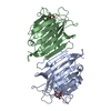 2arbC 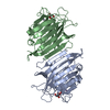 2areC  2auyC 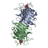 1q8oS C: citing same article ( S: Starting model for refinement |
|---|---|
| Similar structure data |


-Links
 PDBj
PDBj

- Assembly
| Deposited unit | 
| ||||||||
|---|---|---|---|---|---|---|---|---|---|
| 1 |
| ||||||||
| Unit cell |
| ||||||||
| Details | The two chains A and B in the asymmetric unit together form the lectin dimer |
-Components
-Protein , 1 types, 2 molecules AB
| #1: Protein | / PAL Mass: 27575.326 Da / Num. of mol.: 2 / Source method: isolated from a natural source / Source: (natural) Pterocarpus angolensis (plant) / Tissue: seed / References: UniProt: Q8GSD2 |
|---|
-Sugars , 3 types, 3 molecules 
| #2: Polysaccharide | alpha-D-mannopyranose-(1-3)-alpha-D-mannopyranose / 3alpha-alpha-mannobiose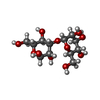  , Oligosaccharide / Class: Metabolism / Mass: 342.297 Da / Num. of mol.: 1 , Oligosaccharide / Class: Metabolism / Mass: 342.297 Da / Num. of mol.: 1Source method: isolated from a genetically manipulated source Details: oligosaccharide / References: 3alpha-alpha-mannobiose |
|---|---|
| #3: Polysaccharide | beta-D-galactopyranose-(1-4)-2-acetamido-2-deoxy-beta-D-glucopyranose-(1-2)-alpha-D-mannopyranose- ...beta-D-galactopyranose-(1-4)-2-acetamido-2-deoxy-beta-D-glucopyranose-(1-2)-alpha-D-mannopyranose-(1-3)-[2-acetamido-2-deoxy-beta-D-glucopyranose-(1-2)-alpha-D-mannopyranose-(1-6)]beta-D-mannopyranose-(1-4)-2-acetamido-2-deoxy-alpha-D-glucopyranose / Mass: 1276.157 Da / Num. of mol.: 1 Source method: isolated from a genetically manipulated source |
| #6: Sugar | ChemComp-NAG / N-Acetylglucosamine Type: D-saccharide, beta linking / Mass: 221.208 Da / Num. of mol.: 1 Type: D-saccharide, beta linking / Mass: 221.208 Da / Num. of mol.: 1Source method: isolated from a genetically manipulated source Formula: C8H15NO6 |
-Non-polymers , 3 types, 214 molecules 


| #4: Chemical |  Mass: 54.938 Da / Num. of mol.: 2 / Source method: obtained synthetically / Formula: Mn Mass: 54.938 Da / Num. of mol.: 2 / Source method: obtained synthetically / Formula: Mn#5: Chemical |  Mass: 40.078 Da / Num. of mol.: 2 / Source method: obtained synthetically / Formula: Ca Mass: 40.078 Da / Num. of mol.: 2 / Source method: obtained synthetically / Formula: Ca#7: Water | ChemComp-HOH / | WaterMass: 18.015 Da / Num. of mol.: 210 / Source method: isolated from a natural source / Formula: H2O |
|---|
-Experimental details
-Experiment
| Experiment | Method: X-RAY DIFFRACTION / Number of used crystals: 1 |
|---|
- Sample preparation
| Crystal | Density Matthews: 2.7 Å3/Da / Density % sol: 53.2 % |
|---|---|
| Crystal grow | Temperature: 293 K / Method: vapor diffusion, hanging drop / pH: 6.5 Details: Na-cacodylate, CaCl2, PEG-8000, Man(a1-3)Man, pH 6.5, VAPOR DIFFUSION, HANGING DROP, temperature 293K |
-Data collection
| Diffraction | Mean temperature: 293 K |
|---|---|
| Diffraction source | Source: SYNCHROTRON / Site: ESRF  / Beamline: ID14-1 / Wavelength: 0.934 Å / Beamline: ID14-1 / Wavelength: 0.934 Å |
| Detector | Type: ADSC QUANTUM 4 / Detector: CCD / Date: Oct 29, 2004 |
| Radiation | Protocol: SINGLE WAVELENGTH / Monochromatic (M) / Laue (L): M / Scattering type: x-ray |
| Radiation wavelength | Wavelength: 0.934 Å / Relative weight: 1 |
| Reflection | Resolution: 2→15 Å / Num. all: 34598 / Num. obs: 34598 / % possible obs: 85.8 % / Observed criterion σ(F): 0 / Observed criterion σ(I): 0 / Redundancy: 3.57 % / Rmerge(I) obs: 0.054 / Net I/σ(I): 12 |
| Reflection shell | Resolution: 2→2.07 Å / Rmerge(I) obs: 0.362 / % possible all: 82.7 |
- Processing
| Software |
| ||||||||||||||||||||
|---|---|---|---|---|---|---|---|---|---|---|---|---|---|---|---|---|---|---|---|---|---|
| Refinement | Method to determine structure: MOLECULAR REPLACEMENT Starting model: PDB entry 1Q8O Resolution: 2→15 Å / Cross valid method: THROUGHOUT / σ(F): 0 / Stereochemistry target values: Engh & Huber
| ||||||||||||||||||||
| Refinement step | Cycle: LAST / Resolution: 2→15 Å
| ||||||||||||||||||||
| Refine LS restraints |
|
